BM 1041:03 Lecture Notes - Lecture 27: Abductor Pollicis Brevis Muscle, Flexor Pollicis Brevis Muscle, Opponens Pollicis Muscle

23 May 2018
School
Department
Course
Professor

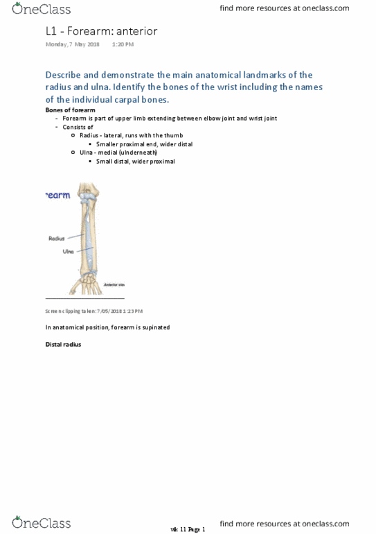
Identify the bones of the hand including the names of the individual carpal bones,
metacarpals and the proximal, middle and distal phalanges.
Screen clipping taken: 15/05/2018 7:33 PM
Hand is region of upper limb distal to wrist joint
-
Wrist/carpus (8) - some lovers try positions that they can't handle
○
Screen clipping taken: 15/05/2018 7:35 PM
Consist of base, shaft/body and head
▪
Bases articulate with carpals and with each other
▪
Heads articulate with proximal phalanges of digits
▪
Heads = knuckles on dorsal surface of hand when fingers flexed
▪
Metacarpus (I-V)
○
Thumb - 2 phalanges (prox and distal)
▪
4 fingers - index, middle, ring, little - have 3 phalanges (prox, middle, distal)
▪
Each has base, shaft and head
▪
Digits - 5 incl thumb
○
3 parts
-
Anterior/palmar
○
Posterior/dorsal
○
2 surfaces
-
Briefly describe the anatomy of the joints of the hand: intercarpal, carpometacarpal
The hand
Tuesday, 15 May 2018
2:55 PM
wk 12 Page 1

Briefly describe the anatomy of the joints of the hand: intercarpal, carpometacarpal
(including the special case of the first CMC joint), metacarpophalangeal, and the
proximal and distal interphalangeal joints. Explain the movements of the fingers and
thumb and identify the muscles responsible for these movements. Name the
function, attachments and nerve supply of these muscles. Differentiate between
those in the forearm and those intrinsic to the hand.
Joints
Screen clipping taken: 15/05/2018 7:38 PM
Joint
Type
Movement
Carpal
Synovial joints between carpal
bones
-
Share common articular cavity
-
Joint capsule reinforced by
ligaments
-
Although movement limited,
contribute to positioning of hand
in wrist movement
-
Carpometac
arpal
5
-
Between metacarpals and distal
row of carpal bones
-
Wide range of mobility to
thumb, unlike rest of digits
•
Flexion, ext, ab, adduction,
rotation, circumduction
•
Saddle jt - between MC I and
trapezius
-
Less mobile
•
Only limited gliding
movements
•
Plane jts - MC II-V
-
Screen clipping taken: 15/05/2018 7:45 PM
Screen clipping taken: 15/05/2018 7:42 PM
So movement of thumb at right angles to other digits
○
Thumb positioned at right angles to orientation of other digits
-
Flex/ext in coronal plane
-
Abduction/adduction = sagittal plane
-
wk 12 Page 2

Abduction/adduction = sagittal plane
-
Opposition
-
Metacarpop
halangeal
Between distal heads of
metacarpals and proximal
phalanges of digits
-
Flex/ext, ab/adduction,
circumduction, limited
rotation
•
Condylar joints
-
Screen clipping taken: 15/05/2018 7:41 PM
Flex and ext in sagittal plane
-
Coronal plane
○
Abduction = spreading fingers away from midline (through middle
finger)
-
Coronal plane
○
Adduction - towards middle finger
-
Interphalang
eal (DIP/PIP)
Hinge joints
-
Mainly flexion and ext
-
Screen clipping taken: 15/05/2018 7:41 PM
Flex and ext in sagittal plane
-
Intrinsic muscles (in hand only)
wk 12 Page 3
Document Summary
Identify the bones of the hand including the names of the individual carpal bones, metacarpals and the proximal, middle and distal phalanges. Hand is region of upper limb distal to wrist joint. Wrist/carpus (8) - some lovers try positions that they can"t handle. Bases articulate with carpals and with each other. Heads = knuckles on dorsal surface of hand when fingers flexed. 4 fingers - index, middle, ring, little - have 3 phalanges (prox, middle, distal) Briefly describe the anatomy of the joints of the hand: intercarpal, carpometacarpal wk 12 page 1. Briefly describe the anatomy of the joints of the hand: intercarpal, carpometacarpal (including the special case of the first cmc joint), metacarpophalangeal, and the proximal and distal interphalangeal joints. Explain the movements of the fingers and thumb and identify the muscles responsible for these movements. Name the function, attachments and nerve supply of these muscles. Differentiate between those in the forearm and those intrinsic to the hand.