BIOC 2300 Lecture Notes - Lecture 6: Amide, Steric Effects, Ramachandran Plot

3 May 2018
School
Department
Course
Professor

Protein Structure
January 18th, 2016
There are four levels of protein structure:
• Proteins are usually defined as polypeptide chains containing over ~50 residues (>5 kDa)
• Their size and complexity requires a hierarchial framework
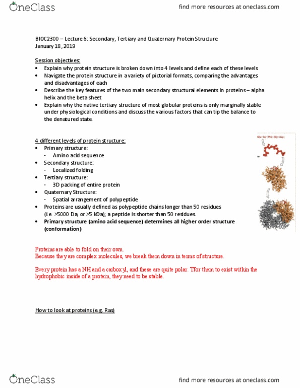
• Primary structure (aa sequence) determines all higher order structure
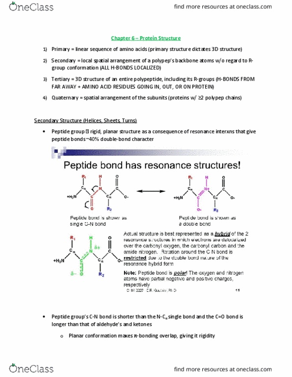
When forming a peptide bond between two amino acids:
• Has a partial double bond character
o Rigid and planar
o Trans configuration (R on opposite sides)
o Uncharged but polar
▪ Made by combination of two charged groups
• Creates a water molecule condensation reaction
• Therefore, creates a stable primary structure
Peptide bonds limits conformations flexibility:
• 3 bonds per amino acid residue
• each amino acid residue alone the peptide backbone contributes 3 bonds ( N-C, C-C, C-
N)
• the peptide bond is planar and roatation about the other two is constrained by steric
hindrance
• this limits possible conformations as seen in a Ramachandran plot **see diagram
• Two secondary structures are commonly found in proteins as an alpha-helix or beta-
pleated sheet
o Alpha helix is stabilized by intra-chain H bonds between backbone amide and
carbonyl groups four residues apart
▪ One complete turn = 0.54 nm = 3.6 residues
▪ R groups project outwards
▪ Most stable in the protein interior
▪ Does not usually contain proline
▪ Folds/unfolds cooperatively
o Beta- sheet is an extended zigzag conformation with H bonds between chains and
R groups project from both faces
▪ May be parallel or antiparallel
▪ Beta-bends often occur where the polypeptide chain makes a 180 degree
turn at protein surface
o The 20 amino acids have different propensities to form either a beta-pleated sheet
or alpha helix
▪ Some are found in one or the other or both
find more resources at oneclass.com
find more resources at oneclass.com
Document Summary
There are four levels of protein structure: proteins are usually defined as polypeptide chains containing over ~50 residues (>5 kda, their size and complexity requires a hierarchial framework, primary structure (aa sequence) determines all higher order structure. Conformational diseases: alzheimer disease, amyloid peptide (1-40) in alpha helix beta-amyloid fibril, prions (cjd, mad cow , undergoes change with more beta sheets. Supersecondary structure: motifs and domains: protein motifs are small regions with a defined sequence or structure that often serves a common function in different proteins, ex. Ef hand calcium ion binding motif: protein domains are sub-regions of single polypeptide chains that can fold and function independently (sometimes correlated with exons, ex. Conservative change amino acid mutates to a similar amino acid. Non conservative change amino acid mutates into a very different amino acid. Molten globule native conformation: energy decreases as the structure becomes more complex to discrete folding intermediates.