BIOL 112 Lecture Notes - Lecture 4: Beta Sheet, Disulfide, Protein Folding

15 Jun 2018
School
Department
Course
Professor

4
BIOL 112: PROTEINS 2 (TERTIARY STRUCTURES)
Primary Structure: first protein, sequence of AA, peptide linkage (300 AA → helix or pleated
sheet)
Secondary Structure: alpha-helix, beta pleated sheet
Tertiary Structure: folding assembles the protein into a specific shape, side chain interactions
determine tertiary structure, H-bond and carbonyl group in the backbone, hydrophobic
interactions + van der waals (pic in slide)
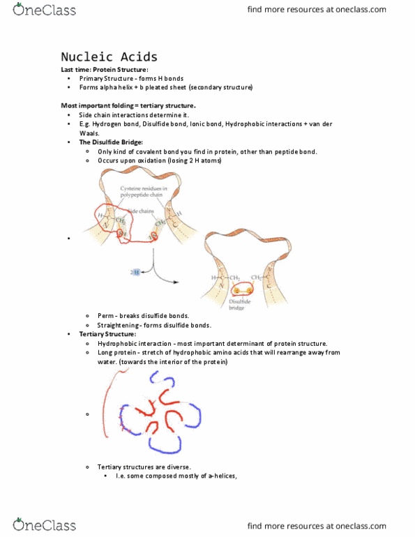
- Disulfide Bridge: Cysteine residues in polypeptide chain, side chains (SH), form covalent
bond called disulfide bridge → which stabilizes the protein
o (e.g. breaking and reforming when getting a perm (adds a chemical to break the
alpha-helixes in your hair and in the end, the disulfide bridge is reformed)
- Hydrophobic Interaction:
o Protein stretches of hydrophobic amino acids (red) will rearrange way from
water (hydrophilic – blue)
▪ Hydrophobic AA are on the interior and Hydrophilic AA are on the
exterior, crucial for protein folding
- Coiled Coils:
o Keratin in your hair → made up of alpha-helixes that form ropes that wind
around each other because they have hydrophobic AA at EVERY 4TH POSITION (1
turn = 3.6 AA) giving rise to the stable structure
The larger the protein sheets, the greater the diversity of alpha helixes and beta pleated sheets
+ disulfide bridges in tertiary folding
Proteins:
- Might hae to for a dier if ca’t fuctio o its o
o E.g. cro protein, polypeptide
o E.g. hemoglobin, a tetramer – 4 different polypeptides that fold independently
but not a functional protein that carries oxygen, they come together to form a
hemoglobin molecule (quaternary structure)
If there is a mutation amongst the AA sequence, they can cause residual effects (e.g. red blood
cells → normal vs. sickled)
Ribonuclease protein folded → disulfide bonds form + hydrogen bonds form
Ribonuclease protein unfolded → disulfide bonds are broken + hydrogen bonds are broken
(unfunctional RNA)
REVERSIBLE REACTION ↑ (ideal circumstances) HOWEVER …
find more resources at oneclass.com
find more resources at oneclass.com


27
BIOL 112 Full Course Notes
Verified Note
27 documents
Document Summary
Primary structure: first protein, sequence of aa, peptide linkage (300 aa helix or pleated sheet) Tertiary structure: folding assembles the protein into a specific shape, side chain interactions determine tertiary structure, h-bond and carbonyl group in the backbone, hydrophobic interactions + van der waals (pic in slide) Hydrophobic interaction: protein stretches of hydrophobic amino acids (red) will rearrange way from water (hydrophilic blue, hydrophobic aa are on the interior and hydrophilic aa are on the exterior, crucial for protein folding. The larger the protein sheets, the greater the diversity of alpha helixes and beta pleated sheets. If there is a mutation amongst the aa sequence, they can cause residual effects (e. g. red blood cells normal vs. sickled) Ribonuclease protein folded disulfide bonds form + hydrogen bonds form. Ribonuclease protein unfolded disulfide bonds are broken + hydrogen bonds are broken (unfunctional rna)