BIOL 201 Lecture Notes - Lecture 12: Cdc42, Profilin, Treadmilling


Leading edge of a cell forms a branched actin meshwork: this is the focus of
what we will see today
•
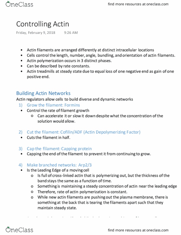
Grow the filament: formins that allow growth faster or slower than
ambient concentrations will allow, gating of growth
1.
Cut the filament: cofilin that disassembles actin network
2.
Cap the filament: capping protein
3.
Make the branched networks: Arp2/3
4.
Cross-link the filaments: ex. α-actinin that forms parallel or antiparallel
arrays of actin
5.
Allow cells to build a diverse and dynamic actin network
•
Function as a dynamic team that coordinates leading edge actin network
•
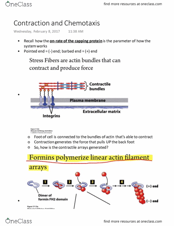
Actin Regulators
Have two actin filament binding domains
•
In fimbrin the actin binding domains are on same
polypeptide chain, alpha actinin has 2 domains that
dimerize, spectrin filaments (cell cortex) that form
alpha and beta heterodimer, filamin is flexible and
contributes to mechanical integrity of overall structure
•
Spacing of actin filaments varies depedning on which
linker is being used
•
Cross-linkers have 2 opposing filament binding domains
Full of crosslinked actin
•
Leading edge is moving out and pushing plasma membrane forward, but
width of actin band remains the same
•
Rate of actin polymerization is also constant, something must be
maintaining relative concentration of actin monomer at the edge
•
The Leading Edge of a moving cell is a branched actin network
Uses actin machinery as part of life cycle life cycle
•
Build pile of actin filaments behind them to move through cytoplasm ("comet
tail") and rocket themselves out of plasma membrane and into neighbouring
cells
•
When bacteria in solution of actin monomers it will behave the same way, and
by altering conditions of medium you can deconstruct the machinery of a motile
actin network
•
Nucleation is rate limiting step!!
○
Listeria “comet tails” are nucleated by the Arp2/3 complex
•
A pathogenic bacterium hijacks the actin machinery of human cells
Come together in what looks like tip of actin filament
•
Spaced out initially, but must first come together to nucleate actin
filament
○
The Arp2/3 complex must be activated!
•
When actin pushing against some load force, of filament pressed
all the way against membrane, a new monomer must be added in
transient space that arises between filament and membrane
○
Cycle of membrane diffusion and actin polymerization
○
As you experience more force, the velocity can acheive goes down,
and so does critical angle
○
70 degrees provides just enough space for this process to occur at
an optimal rate
○
70° creates optimal angles for pushing out the plasma membrane
•
Has 3 domains: W, A and C
○
WH2 domain binds an actin monomer, A domain binds Arp 2/3
complex that brings its monomers togehter
○
r
Wiskott-Aldrich Syndrome family proteins (WASp) activate Arp2/3
•
Arp2/3 mimics an actin nucleus and nucleates daughter filaments at 70°
angles
Lecture 12: Controlling Actin
February 9, 2018
9:48 AM
Tutorials Page 1
Document Summary
Leading edge of a cell forms a branched actin meshwork: this is the focus of what we will see today. Allow cells to build a diverse and dynamic actin network. Grow the filament: formins that allow growth faster or slower than ambient concentrations will allow, gating of growth. Cut the filament: cofilin that disassembles actin network. Actinin that forms parallel or antiparallel arrays of actin. Function as a dynamic team that coordinates leading edge actin network. In fimbrin the actin binding domains are on same polypeptide chain, alpha actinin has 2 domains that dimerize, spectrin filaments (cell cortex) that form alpha and beta heterodimer, filamin is flexible and contributes to mechanical integrity of overall structure. Spacing of actin filaments varies depedning on which linker is being used. The leading edge of a moving cell is a branched actin network. Leading edge is moving out and pushing plasma membrane forward, but width of actin band remains the same.