ANAT 261 Lecture 11: Lecture 11 – Blood Vessels

18 Feb 2019
School
Department
Course
Professor

October 10th, 2017
Lecture 11 - Blood Vessels
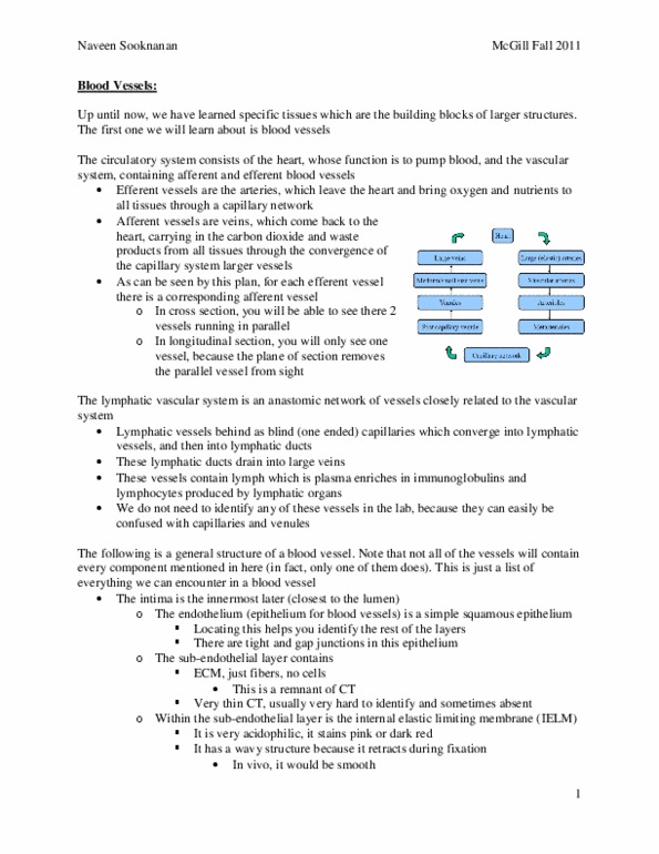
Circulatory System: Composed of the Heart and Vascular System
The Vascular System:
• Efferent Vessels: Arteries leaving the heart to transport
nutrients and oxygen to the tissues. Nutrients and
oxygen diffuse out of the blood vessels and into the
tissues at the level of the capillary network.
o Large arteries: aorta and carotid artery
• Afferent Vessels: Veins returning to the heart, carrying
CO2 and metabolic waste from the tissues
o Large Veins: Superior and Inferior Vena Cava
Cross-Section (aka Transverse Section) of Blood Vessels
• Veins and arteries run in parallel to each other (as seen on
image on the left)
• Consequence: Arteries and veins appear side by side and
running in the same direction when being viewed in cross-
section
o Advantage of looking at blood vessels in cross
section
• Examples:
o Large artery will appear next to a large vein
o Muscular artery will appear next to a muscular vein
o Arteriole will appear next to a venuole
o Metarteriole will appear next to a post-capillary
venuole
• When looking at a cross-section of blood vessels the
smooth muscle cells (of the media layer) will appear in
longitudinal section
• Comparing Arteries and Veins in Cross-Section
o Both veins and arteries may have red blood cells in the lumen (space in the
middle)
o When trying to differentiate between arteries and venules in cross-section, look
at the shape, thickness of the media and whether the structure is collapsed.
o Shape:
▪ Artery: shape will appear round (not collapsed)
• Artery keeps its shape because the Media layer (M) is very large,
you have many layers of smooth muscle cells
▪ Vein: Shape is Oval (tends to be collapsed) and may appear much larger
than this one (in the femur, muscular artery and vein example)
find more resources at oneclass.com
find more resources at oneclass.com

October 10th, 2017
• Has a very thin Media Layer (M) which contains much fewer
smooth muscle cells than an artery of equal size and it and has an
adventitia (dense irregular CT) that is very large compared to the
Media Layer.
▪ Why the artery keeps its shape but the vein doesn’t:
• The wall of the vein (Media Layer) is very thin (compared to the
artery). Therefore, when you fix the tissue with formaldehyde you
will see that the arteries keep a nice shape due to its large Media
of smooth muscle cells (supporting it) and the Vein tends to be
collapsed due to its very thin Media.
Longitudinal Section of Blood Vessels
• Runs in same direction as blood vessel (blood vessels run longitudinally along the body)
• When looking at the Media Layer of Blood Vessels in longitudinal section, the smooth
muscle cells will appear in cross section
Perfect Longitudinal Section
• In a perfect longitudinal section (cut for example right down the middle of the artery
and the vein) you will still see the Artery right next to the Vein, except now you will see
them in a longitudinal orientation
find more resources at oneclass.com
find more resources at oneclass.com

October 10th, 2017
• Note: Structures (blood vessels) don’t always appear straight, they may appear
curved/distorted like in the example below (due to plane of section)
• Muscular Artery (from longitudinal section shown below):
o Longitudinal section
o Lumen has some RBC inside
o Media Layer (M) is still very thick
• Muscular Vein
o Practically has no Media, you only Adventitia
o Adventitia of the vein is fused with the adventitia of the artery
o Lumen appears red in this slide because of red blood cells
• Comparing Arteries and Veins in longitudinal Section (same way as you would in cross
section):
Longitudinal Sections may have ONLY an artery or a vein
• You lose your point of reference, there’s no artery to compare it to so it’s harder to see
that it’s a vein
• You can still identify whether it’s an artery or a vein using the same rules for comparing
arteries to vein: Look at (1) shape, (2) Media Layer, and (3) whether or not it’s collapsed
o In longitudinal section, it’s hard to identify whether it’s collapsed so focus on
Media Layer
o Ex (Slide below): There is practically no visible media, all you really see are
Endothelial cells covering the surface of the vein
find more resources at oneclass.com
find more resources at oneclass.com
Document Summary
Circulatory system: composed of the heart and vascular system. The vascular system: efferent vessels: arteries leaving the heart to transport nutrients and oxygen to the tissues. Nutrients and oxygen diffuse out of the blood vessels and into the tissues at the level of the capillary network: large arteries: aorta and carotid artery, afferent vessels: veins returning to the heart, carrying. Co2 and metabolic waste from the tissues: large veins: superior and inferior vena cava. Media layer: why the artery keeps its shape but the vein doesn"t, the wall of the vein (media layer) is very thin (compared to the artery). In a perfect longitudinal section (cut for example right down the middle of the artery and the vein) you will still see the artery right next to the vein, except now you will see them in a longitudinal orientation. In longitudinal section, it"s hard to identify whether it"s collapsed so focus on.