BIOC 212 Lecture Notes - Lecture 9: Glycoprotein, Calreticulin, Mannose

28 May 2018
School
Department
Course
Professor

5- Membrane Proteins
!
Signal Anchors
• Signal anchor: signal sequence with long hydrophobic region that also becomes
the TM domain (longer, 18-24aa)
o Signal sequence is long enough to itself become a TM domain
o Not cleaved off, can be in middle of protein as well as at the N-terminus
• Not removed given that serve as a TM helix
o Signal anchors start translocation, then are membrane integrated
• Signal is recognized by SRP, brought to membrane and associate to
translocon
• Move sideways and integrated in membrane
• TM signal anchor can be recognized by the translocon in different orientations
o Either way still associate sideways
• Charges in protein sequence next to each side of the signal anchor are recognized
by translocon and determine the orientation
o Orientation depends on charges on each side of hydrophobic region of
signal anchor
o Positive charges in cytosol & negative charges in lumen
• Evolved this way, no biological reason for it
o The translocon recognizes the charge pattern and keep the signal anchor in
that orientation as it is being integrated into the membrane
• In one situation, have the N-terminus in the lumen and the ribosome continues
translating the rest in the cytosol
o As with previous conditions
• In the other, have the N-terminus in the cytosol, and the ribosome pushes the rest
of the polypeptide into the lumen
Multi-Pass TM Proteins
• Combinations of signal anchors & TM helices cause alternating orientation of multi-
pass TM proteins
o Might start with a signal anchor, in this example giving the N-terminus in the
lumen, and then have alternating TM helices and signal anchors that
eventually stitch the protein into the membrane
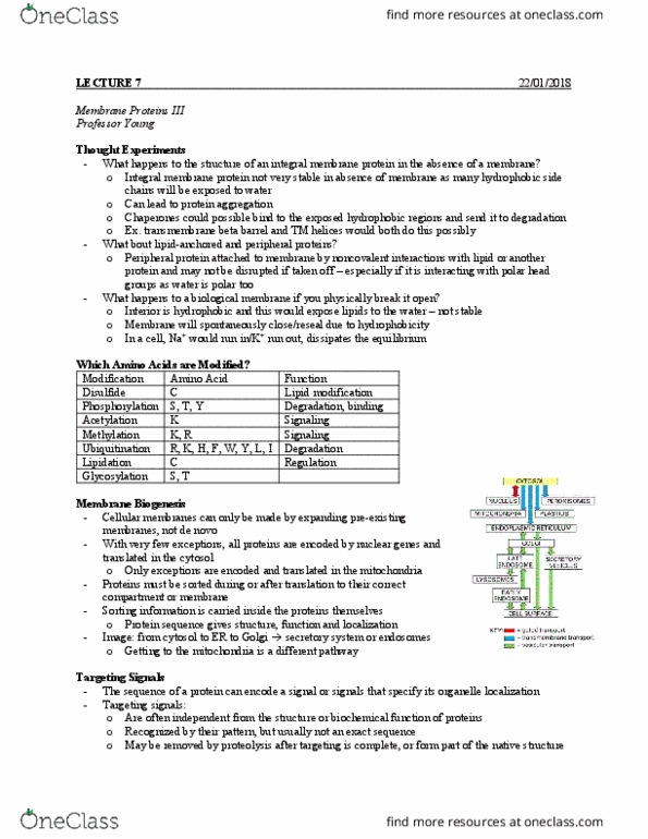
• Topology (TM organization) of secretory pathway proteins can often be predicted
from:
o Hydrophobicity – Number of TM helices
o Charge distribution – Orientation in membrane
• Look at the charges on each side of the hydrophobic regions, can predict
which way around those helices go

o Can then predict other modifications – Disulfide bonds, glycosylation,
phosphorylation, ubiquitination
• Once know the orientation of the segments in the membrane, can look at
where modifications should go
N-Linked Glycosylation
• Most secretory proteins have oligosaccharides (glycans) covalently attached
1. Help stabilize the native state
• Carbohydrates are very polar, so they want to stay outside the protein
and keep hydrophobic segments inside
2. Protect against proteases by covering with sugars
3. Function in cell surface signaling
• Help protein determine when they are folding/folded?
• Oligosaccharides are a chain a small monosaccharides
• N-linked glycosylation on Asn side chain in context of Asn-X-Ser/Thr motif
o Always on the nitrogen atom on an asparagine (N)
• A glutamine cannot substitute
• Enzyme is very specific for asparagine
o Motif recognized made of asparagine, any amino acid and then Ser or Thr
o The same glycan chain is always attached at ER
• Mixture of different sugars
• Mostly made of mannose, and has a glucose at the end (3)
Asparagine (N)
• N-linked glycosylation on Asn side chain amide
o Gln is not recognized by Oligosaccharyl Transferase (OST)
o The pocket on the enzyme that recognizes Asn is too short for Gln (extra
CH2)
• Most N-x-S/T motifs in the lumen are modified, depending on accessibility to the
enzyme OST
• Glycans can be modified after addition, but are not removed until protein is
degraded
o Once have glycan on, can change the sugars but cannot entirely remove
them until degrade the protein
Glycosylation Process
• Oligosaccharides are synthesized attached to dolichol phosphate, a specialized
lipid in the ER
o Glycan chain (always the same) is synthesized separately from the protein
o Pre-assembled onto special lipids called dolichol phosphate
• OST will transfer the glycan from the special lipid to the protein during
translocation