BIOC 212 Lecture Notes - Lecture 22: Occludin, Cytoskeleton, Calcification

29 May 2018
School
Department
Course
Professor

Cell Junctions & Cell Adhesion II
!
Dimension in Life Sciences
• 1mm = 10-3 m
• 1um = 10-6 m
• 1nm = 10-9 m
• 1mm scale seen with naked eye
• 10-100um and smaller not seen with naked eye; eukaryotic cells
• Viruses around 100nm
• Proteins, enzymes, etc. between 5-10nm
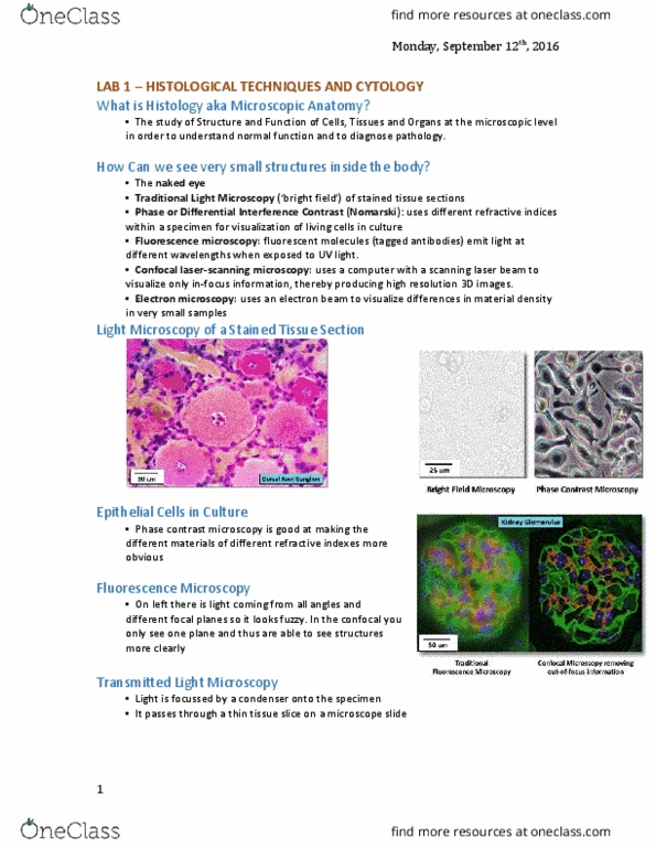
• Light microscopy: range/solution determined by wavelength of light
o 400-700nm = range of visible light
o Explains why cannot be used to see objects smaller than 400nm
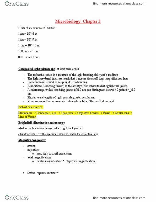
• Electron microscope: uses electron as medium to visualize specimens
o The faster you accelerate the electrons, the smaller is the wavelength
• High power on electron source causes them to fly faster
o Can visualize objects that are in the < 1nm range
Electron Microscopy (Transmission)
• Light microscope:
o Light source with series of condenser lenses that focuses light beam on
biological specimen
o Lenses also provide with magnification
o Use eye or camera to visualize
• Electron microscope:
o Uses magnetic rings to focus electron beam
o Source of electrons = cathode at the top
• Acceleration through nearby anode
o Electron beam travels down the tube
o High power (100 000V) to get small wavelength (0.004nm)
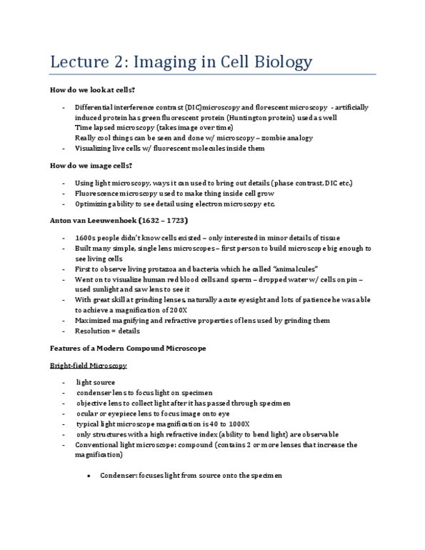
• Biological specimen must be very thin
o Electrons would get absorbed by thicker specimen
• Electrons become scattered by collisions with air molecules
o Ultralow vacuum necessary
o If air was present in the tube (2-3m long), as electron beam travelled through,
the electrons would be absorbed
• Magnetic coils focus the beams (like glass lenses in a light microscope)
• Staining of specimen with electron dense material
o i.e. osmium tetroxide --binds to lipid bilayers & proteins
o Visualization