BIOLOGY 2B03 Lecture Notes - Lecture 4: Transmission Electron Microscopy, Scanning Electron Microscope, Optical Microscope

27 Sep 2016
School
Department
Course
Professor

Document Summary
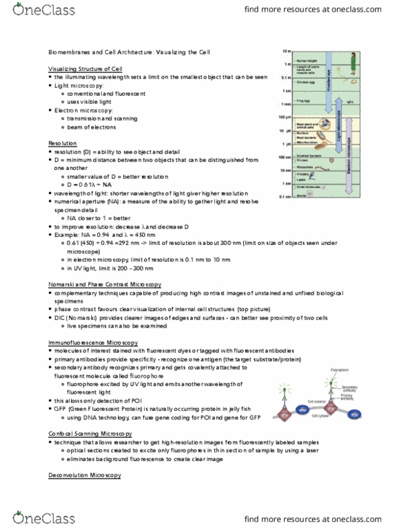
Cytoplasm: cytoskeleton, and organelles of the eukaryotic cell (organelles) Illuminating wavelength sets a limit on the smallest object that can be seen. Light microscopy: conventional and fluorescent- uses visible light. Electron microscopy: transmission and scanning beam of electrons. Light microscope: plant and animal cells, nucleus, bacteria, mitochondria (1 m-100 m) Resolution the ability to distinguish between two very closely positioned objects. D = resolution or limit of resolution; minimum distance to distinguish two objects. Wavelength, shorter wavelengths of light give higher resolution. Na numerical aperature, angle of the light entering the objective and the ability of a medium to bend the light. Light microscopy: visible light ( = 400nm to 700nm) Electron microscopy: beam of electrons, shorter therefore much lower d = better resolution. Brightfield microscopy simplest of all the optical microscopy illumination: observed through white light; involves the use of an organism mounted to a glass microscope slide. Darkness or brightness depends on refractive index of that region.