HTHSCI 1LL3 Lecture Notes - Lecture 17: Polysaccharide, Lacteal, Jaundice

29 Mar 2017
School
Department
Course
Professor
Document Summary
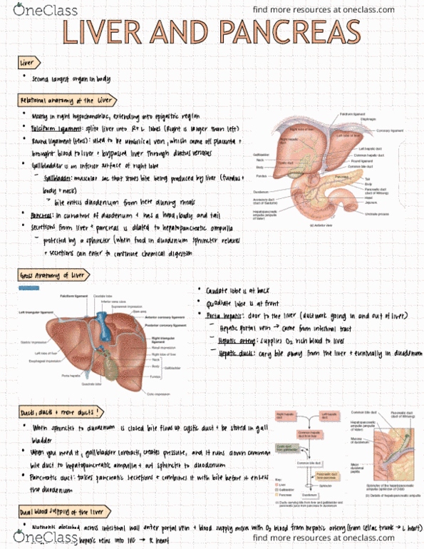
Double fold of peritoneum that divides the l and r lobes. Ligamentum teres = lies within the folds of the falciform ligament (remnant of umbilical vein) Merge in the middle of the porta hepatis to form the common hepatic duct. Liver is connected to duodenum by a set of ductwork. Green sack that stores bile when body is not digesting food in duodenum. Opening of this tube is protected by the sphincter. When there"s food present in the duodenum, sphincter relaxes then digestive secretions from pancreas and liver can now make their way to the duodenum. Gross anatomy of the liver (refer to slide) R lobe = larger than left; subdivided into inferior & posterior lobe; l lobe = smaller. Quadrate lobe = towards the front square. Where duct work/blood vessels (that goes in/out of liver) sits. Hepatic portal vein = branches goes to l and r lobes.