PSYCH 1XX3 Lecture Notes - Lecture 8: Reticular Formation, Midbrain Tectum, Brain Injury


Neuroscience II
The Structure of the Brain
Terminology
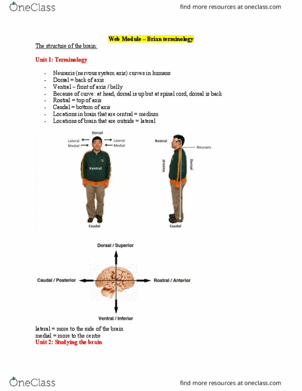
- Nervous system Axis “Neuraxis”
o Curves into the head
- Dorsal: refers the back of the axis
o Also means up
- Ventral: in front of the axis or “to the
belly”
- Rostral: towards the top of the axis
- Caudal: towards the bottom of the axis
- Medial: towards the inside of the brain
- Lateral: towards the outside of the
brain
Studying the Brain
Lesion studies
- Special techniques exist for studying the structure and function of the brain
- Most information about the nervous system comes form animal
- Lesion Studies: conduct studies on neurological patients who have focal brain lesions
o Lesion: any abnormal damage or change in the tissue of an organism
- Brain injury links brain anatomy with behavioural deficits Ex Phineas Gage
- Lesion studies must be specific to target the function of a brain region
o Advantage: A direct measure of a brain structure’s function
o Disadvantage: hard to selectively target regions and draw conclusion
o Solution: Specific brain lesion can be studied in animal models
Stimulation and Single Cell Recording
- Targeted electrical stimulation can also provide insight on brain function
o Electrically stimulate a region of the brain and see what it does to behaviour
o Used extensively by Dr. Wilder Penfeild
- Single cell recording reveals the function of individual neurons
- Disadvantage: limited to only a single neuron
Structural Neuroimaging
- Large-scale brain structures are studied using neuroimaging techniques
- CT Scans: produce structural slices of the brain using X rays
o Relatively quick and inexpensive
- MRIs provide higher resolution images of the brain
o Magnetic fields are generated which align hydrogen atoms
o Helps locate tissue
o Produce detailed pictures of organs, soft tissues, bone and virtually all other internal body
structures.
find more resources at oneclass.com
find more resources at oneclass.com

Functional Neuroimaging
- PET scans display the functional role of brain structures
o Radioactive tracer is injected, and more active brain regions will use more
metabolic resources
o Disadvantage: invasive procedure
- fMRI is a less invasive functional neuroimaging technique
o Produces a clear image without using a radioactive tracer
o Works by measuring blood oxygen dependent signal, and uses many of the same principles as
the MRI
o Works almost the same as a PET scan but with oxygen and its presence in metabolic processes
- Disadvantage: fMRIs provide temporally imprecise mapping of brain function, since oxygen spikes a
few seconds later
o Not the best method for precise timing of brain activation and function
- EEGs display the activity from specific populations of neurons
o Wearing a cap
o By averaging EEGs over a period of time, the noise can be balanced out
o EEG and ERP signals can be highly informative markers with precise temporal resolution on the
order of milliseconds
The Brain Regions – Hindbrain and Midbrain
Hindbrain
- Connects the brain to the spinal cord
- Consists of the Medulla, Pons, Reticular formation, and
the cerebellum
- Are the oldest parts of the brain; primarily involves in regulating vital bodily functions
Brain Region
Function
Medulla
Regulates breathing, digestion and heart rate
Pons
Relay information about movement; auditory
perception and emotional processing. Sleep eye
movement
Reticular Formation
Roles in arousal and motivation, circadian rhythm
and posture and balance, sleep-wake cycle
Cerebellum
Facilitates coordinated movement
Midbrain
- Processes perception, arousal, and motor control
- Consists of the Tectum and the Tegmentum
- Tectum has two regions involved in perception and action
o Superior Colliculi: involved in eye movement and visual reflexes
o Inferior Colliculi: involved in auditory integration
- Tegmentum
o Red Nucleus: contributes to motor control
o Substantia Nigra: role in reward-related behaviours through the release of dopamine
find more resources at oneclass.com
find more resources at oneclass.com
Document Summary
Nervous system axis neuraxis : curves into the head. Dorsal: refers the back of the axis: also means up. Ventral: in front of the axis or to the belly . Rostral: towards the top of the axis. Caudal: towards the bottom of the axis. Medial: towards the inside of the brain. Lateral: towards the outside of the brain. Special techniques exist for studying the structure and function of the brain. Most information about the nervous system comes form animal. Lesion studies: conduct studies on neurological patients who have focal brain lesions: lesion: any abnormal damage or change in the tissue of an organism. Brain injury links brain anatomy with behavioural deficits ex phineas gage. Targeted electrical stimulation can also provide insight on brain function: electrically stimulate a region of the brain and see what it does to behaviour, used extensively by dr. wilder penfeild. Single cell recording reveals the function of individual neurons. Disadvantage: limited to only a single neuron.