BPK 142 Lecture Notes - Lecture 7: External Intercostal Muscles, Alveolar Pressure, Bronchus

27 Mar 2020
School
Department
Course
Professor
Document Summary
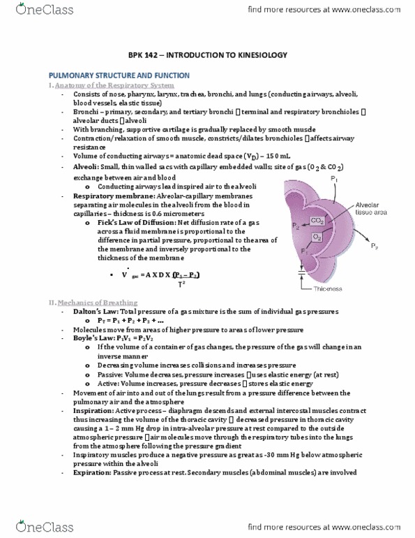
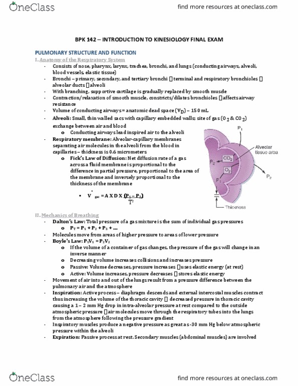
Respiratory system consists of nose, pharynx, larynx, trachea, bronchi, and lungs. Bronchi - primary, secondary, and tertiary bronchi terminal and respiratory bronchioles alveolar ducts alveoli: with branching, supportive cartilage is gradually replaced by smooth muscle. Contraction and relaxation of this smooth muscle constricts or dilates the bronchioles major effects on airway resistance. The conducting airways lead inspired air to the alveoli. Volume of conducting airways = anatomic dead space (vd) - 150 ml. Alveoli: small, thin walled sacs that have capillary beds in their walls; site of gas molecule (o2 & co2) exchange between air and blood. Respiratory membrane: alveolar-capillary membranes that separate the air molecules in the alveoli from the blood in the capillaries - average thickness is 0. 6 micrometers. Evolution gives us 2 cell layers to diffuse to decrease thickness. During exercise: increasing ventilation opens more alveoli thus increasing area. Increase p1 by increasing ventilation and thus the rate of entry of o2 into the lungs.