ZOO 3000 Lecture Notes - Lecture 1: Desmosome, Cadherin, Microvillus

8 Feb 2018
School
Department
Course
Professor
Document Summary
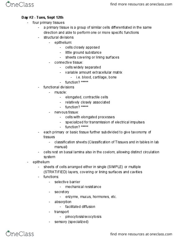
Image: junctional complex: a. electron micrograph of the apical portion of two adjoining epithelial cells of the gastric mucosa, showing the junctional complex. It consists of the zonula occludens (zo), zonula adherens (za), and macula adherens (ma). Diagram showing the distribution of cell junctions in the three cellular domains of columnar epithelial cells. The apical domain with microvilli has been lifted to better illustrate spatial arrangements of junctional complexes within the cell. Diagram of zonula adherens: molecular organization of zonula adherens. Actin filaments of adjacent cells are attached to the e-cadherin catenin complex by - actinin and vinculin. The e-cadherin catenin complex interacts with identical molecules embedded in the plasma membrane of the adjacent cell. Interactions between transmembrane proteins are mediated by calcium ions. Diagram showing three transmembrane proteins involved in the formation of zonula occludens: occludin, claudin, and junctional adhesion molecule (jam).