BIOB11H3 Lecture Notes - Lecture 23: Signal Transduction, Guard Cell, Sea Urchin


Lec 23- Signal Transduction III
Lecture Notes
• Calcium Signaling
• Ca2+ stored in ER, IP3 activates ion channel, triggering Ca2+ release which is a…
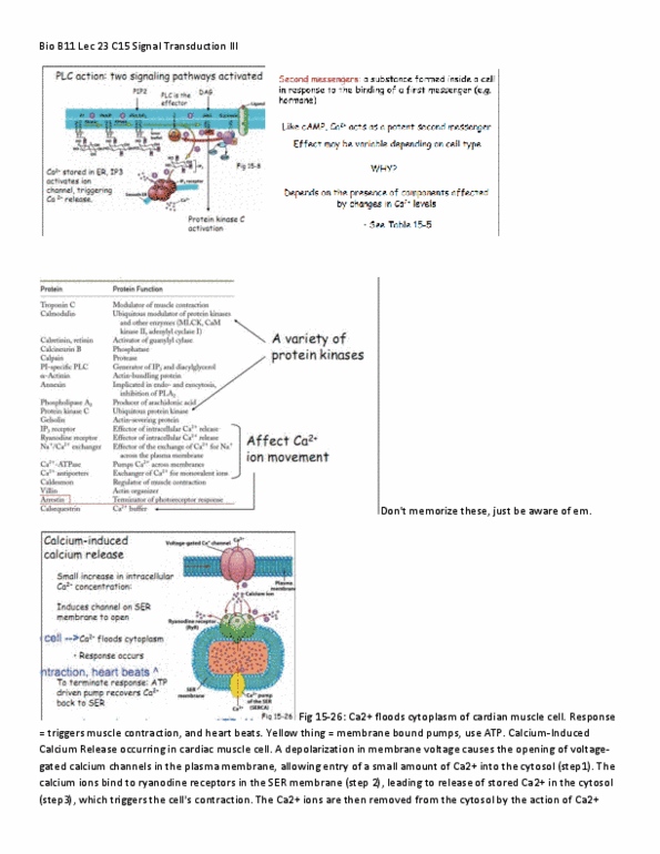
• Second messengers: a substance formed inside a cell in response to the binding of a first
messenger (e.g. hormone)
• Like cAMP, Ca2+ acts a s potent second messenger but effect may be variable depending
on cell type & presence of components affected by changes in Ca2+ levels
• Following are 3 examples of calcium induced calcium release →
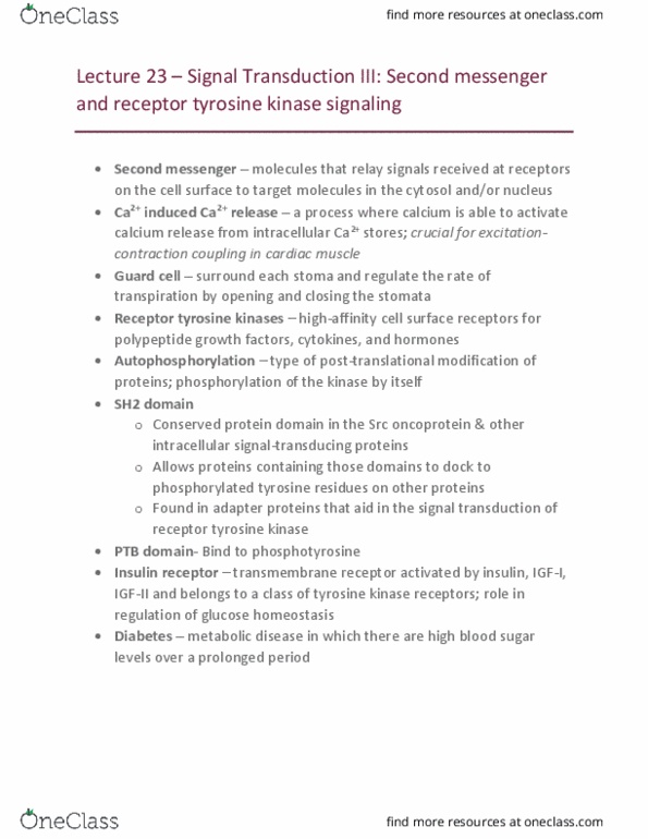
• Fig 15-28: Example #1 –Cardiac muscle
• 1. Depolarization in membrane voltage causes the opening of the voltage-gated calcium
channels in the plasma membrane, allowing entry of small amount of Ca2+ into cytosol
• 2. Calcium ions bind to receptors in SER membrane
• 3. Release of stored Ca2+ into the cytosol which triggers cells contraction
• 4. Calcium ions removed from cytosol by Ca2+ pumps located in SER membrane
• 5. Na+/Ca2+ secondary transport system in plasma membrane also helps transport
• Fig 15-29: Example #2- Sea urchin eggs
• Calcium wave initiated by fertilization drives cell division
• Unfertilized egg was injected w/ calcium-sensitive fluorescence dye, fertilized &
photographed to see rise in Ca2+ as internal calcium released from SER
• Now Ca2+ activates cell division through CDK activation; rapid embryogenesis occurs
• Fig 15-32: Example #3- Guard cells & stomata
• Stomate: pore
• Guard cells: specialized pair that controls stomatal aperature
• Guard cell shape changes to control pore opening
• Stomata are kept open as turgor pressure is kept high within the guard cells, causing them
to bulge outward
• ABA: hormone controlling stomatal pore size as calcium responds to it
o High temp, low humidity causes ABA to be produced
o H2O loss regulated by changes in solute (K+) concentration
• 1. When ABA levels rise, calcium ion channels in the plasma membrane are opened
allowing the influx of Ca2+
• 2. Triggers the release of Ca2+ from internal stores
• 3. Subsequent elevation of intracellular [Ca2+] closes K+ influx channels & opens K+
efflux channels
• 4. These ion movements lead to a drop in internal solute concentration & the osmotic loss
of water
• Receptor Tyrosine Kinases (RTK)
• Two general intracellular STPs; one is G-protein mediated & other uses RTKs
• Fig 15-17: Steps in the activation of an RTK
find more resources at oneclass.com
find more resources at oneclass.com
Document Summary
Depolarization in membrane voltage causes the opening of the voltage-gated calcium channels in the plasma membrane, allowing entry of small amount of ca2+ into cytosol: 2. Calcium ions bind to receptors in ser membrane: 3. Release of stored ca2+ into the cytosol which triggers cells contraction: 4. Calcium ions removed from cytosol by ca2+ pumps located in ser membrane: 5. When aba levels rise, calcium ion channels in the plasma membrane are opened allowing the influx of ca2: 2. Triggers the release of ca2+ from internal stores: 3. Subsequent elevation of intracellular [ca2+] closes k+ influx channels & opens k+ efflux channels: 4. Once bound tyrosine residues on the docking protein are phosphorylated by the receptor. Insulin receptor exists as a tetramer w. 2 extracellular subunits & 2 transmembrane subunits: 1. Conformational change in , kinase activation: 3. Insulin receptor substrate (irs) molecules also phosphorylated (when they bind to receptor. Activation of pi3k enzyme leads to pip3 lipid formation: 2.