BIOC33H3 Lecture Notes - Magnetic Resonance Angiography, Myoglobin, Systolic Geometry
55 views7 pages

25 Mar 2013
School
Department
Course
Professor
Document Summary
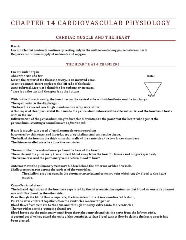
The heart is a four-chambered organ that lies in the mediastinal space in the thorax. The heart is divided by the septum, forming the right and left atrium and the right and left ventricle. The heart is: composed of three layers: endocardium, myocardium, and epicardium, surrounded by a fibroserous sac called the pericardium. The right side of the heart receives blood from the body (via the vena cava) and pumps it to the lungs where it is oxygenated. Blood returns to the left side of the heart (via the pulmonary arteries) and is pumped to the body via the aorta. The coronary circulation provides blood to the myocardium. The right and left coronary arteries are the first branches of the aorta. The conduction system consists of specialized cells that create and transport electrical impulses. These electrical impulses initiate depolarization (contraction) of the myocardium and ultimately a cardiac contraction. The electrical activity of the heart is recorded on the electrocardiogram (ecg).
Get access
Grade+20% off
$8 USD/m$10 USD/m
Billed $96 USD annually

Homework Help
Study Guides
Textbook Solutions
Class Notes
Textbook Notes
Booster Class
40 Verified Answers
Class+
$8 USD/m
Billed $96 USD annually

Homework Help
Study Guides
Textbook Solutions
Class Notes
Textbook Notes
Booster Class
30 Verified Answers
Related Documents
Related Questions
Match the part of the human heart with its function
|
|

