BIO130H1 Lecture Notes - Lecture 19: Optical Tweezers, Intermediate Filament, Electron Microscope


31
BIO130H1 Full Course Notes
Verified Note
31 documents
Document Summary
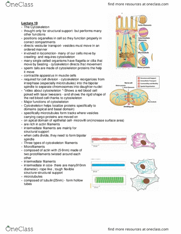
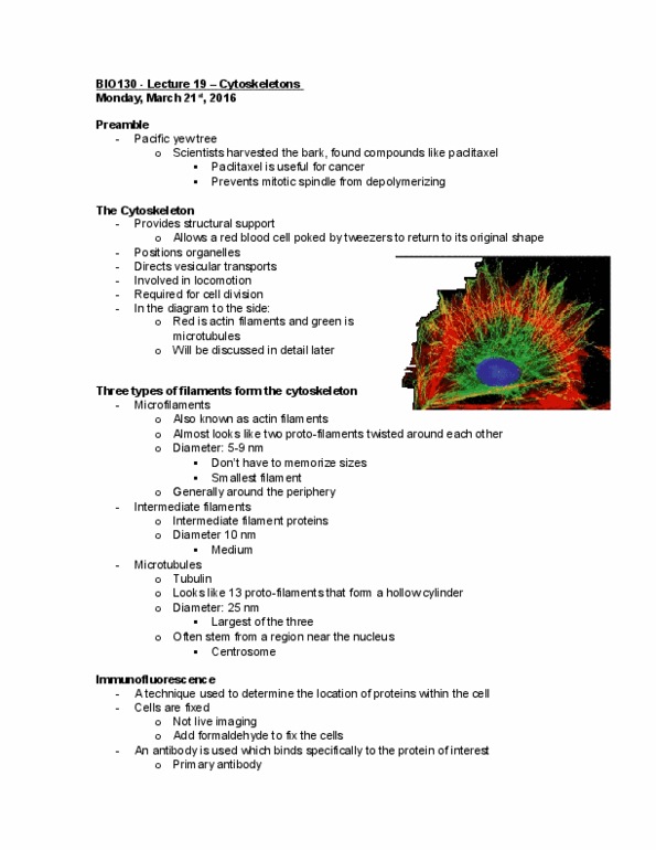
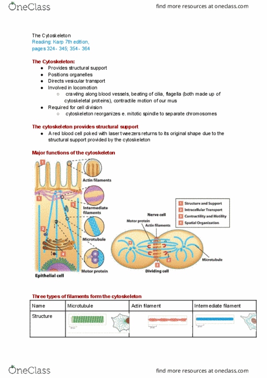
Technique: red blood cell poked with laser tweezers. This is like when the rbc is squeezed through a small crevice. Often found in the periphery of the cell. Often stem from the nucleus area, the centrosome. Appears to be 13 protofilaments to form a hollow center. Technique used to determine location of proteins within cell. An antibody is used which binds specifically to protein of interest. Second antibody binds to first and is covalently tagged with fluorescent molecule. This is used because it"s cheaper and more practical. A fluorescence microscope is used to excite fluorescent molecule and visualize light emitted. Multiple antibodies can be used to fluorescence multiple structures simultaneously. Light microscope has resolution limit due to diffraction (bending of light/waves as it passes) Based on wavelength of light, typically 250 nm. 250x better resolution than light microscope (clearer photo) Light microscopy: can see the flagella easily with fluorescence. Electron microscope allows for viewing cross-sections in detail.