Thursday, March 12, 2008
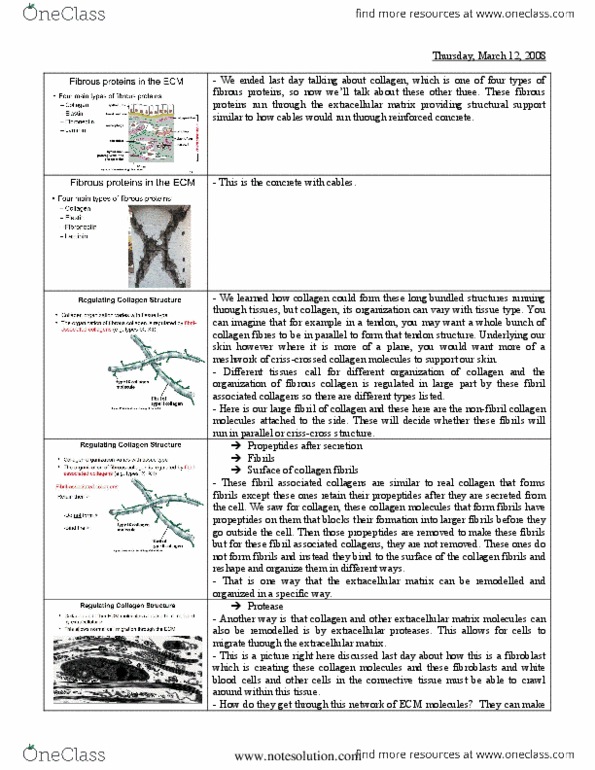
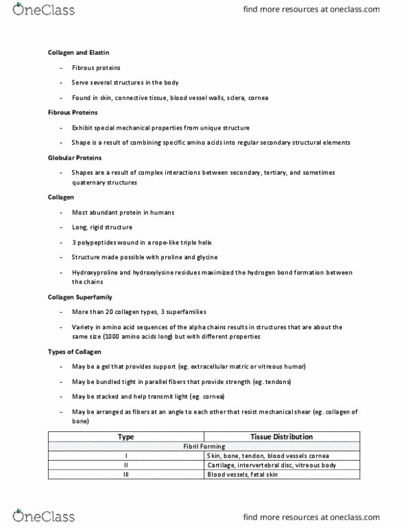
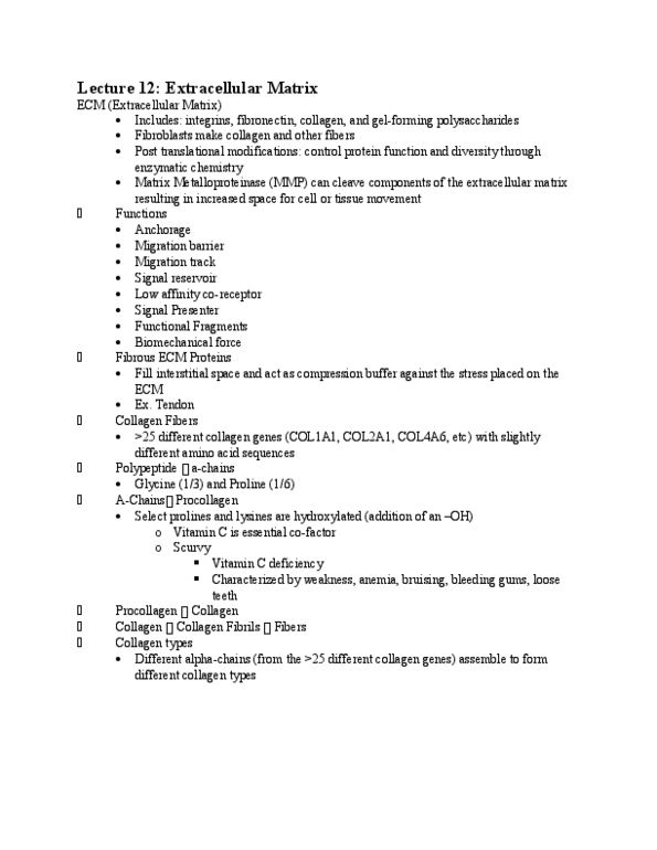
- We ended last day talking about collagen, which is one of four types of
fibrous proteins, so now we’ll talk about these other three. These fibrous
proteins run through the extracellular matrix providing structural support
similar to how cables would run through reinforced concrete.
- This is the concrete with cables.
- We learned how collagen could form these long bundled structures running
through tissues, but collagen, its organization can vary with tissue type. You
can imagine that for example in a tendon, you may want a whole bunch of
collagen fibres to be in parallel to form that tendon structure. Underlying our
skin however where it is more of a plane, you would want more of a
meshwork of criss-crossed collagen molecules to support our skin.
- Different tissues call for different organization of collagen and the
organization of fibrous collagen is regulated in large part by these fibril
associated collagens so there are different types listed.
- Here is our large fibril of collagen and these here are the non-fibril collagen
molecules attached to the side. These will decide whether these fibrils will
run in parallel or criss-cross structure.
Propeptides after secretion
Fibrils
Surface of collagen fibrils
- These fibril associated collagens are similar to real collagen that forms
fibrils except these ones retain their propeptides after they are secreted from
the cell. We saw for collagen, these collagen molecules that form fibrils have
propeptides on them that blocks their formation into larger fibrils before they
go outside the cell. Then those propeptides are removed to make these fibrils
but for these fibril associated collagens, they are not removed. These ones do
not form fibrils and instead they bind to the surface of the collagen fibrils and
reshape and organize them in different ways.
- That is one way that the extracellular matrix can be remodelled and
organized in a specific way.
Protease
- Another way is that collagen and other extracellular matrix molecules can
also be remodelled is by extracellular proteases. This allows for cells to
migrate through the extracellular matrix.
- This is a picture right here discussed last day about how this is a fibroblast
which is creating these collagen molecules and these fibroblasts and white
blood cells and other cells in the connective tissue must be able to crawl
around within this tissue.
- How do they get through this network of ECM molecules? They can make use of proteases to actually cut these molecules, chop them so they can go
through. The action of these protease, this has to be highly regulated so you
should note that these molecules, the proteases are secreted from cells in an
inactive form, and they’re normally secreted along with inhibitors so you
secrete the proteases and also inhibitors of those proteases. Generally you
don’t want these proteases to work except in very specific circumstances. If
you sent out active proteases into the ECM, it would digest the entire ECM
and it would be gone. These inactive molecules are sent out into the
extracellular space and normally their activity is controlled by binding to the
surface of the cell that wants to use them. These inactive molecules will bind
to the surface of the cell & therefore be locally activated & very specifically
controlled. You have very specific digestion of the extracellular matrix where
you need a fibroblast to crawl through it or a white blood cell, etc.
Proteases
- In addition to these cell types, this is also relevant to cancer progression. As
we’ll learn about and as we’re all aware, we will have a primary tumour and
then as cancer progresses, the cells will start to migrate away from that
primary tumour to other sites in the body. To migrate from the primary
tumour, those cells also have to travel through the extracellular matrix. They
also make use of proteases to do that.
- It is important to know about these proteases because there is the potential
to develop drugs to block the ability of cancer cells to travel through the
extracellular matrix so you might be able to slow down the progression of
cancer, stopping them from spreading through the extracellular matrix.
- That is just shown in the cartoon. There is a tumourous cell that is growing
and metastasizing, moving through the extracellular matrix. It is doing that
here because it has these uPA receptors being expressed so therefore it is
recruiting this protease called uPA to the surface of the cell and activating it
there. Now this cell will have active proteases coating its surface so when it
comes into contact with extracellular matrix, it can chop those proteins up
and travel through that extracellular matrix.
- On the other side, there is an inactive form of the protease that is being
expressed, this is competing away the normal protease so most of these
receptors on the surface of the cell are now bound to inactive proteases. You
can imagine in this case, this is a mutant form of the protease that
outcompetes the normal form but you could also imagine that these yellow
molecules could be a drug that could be given to a cancer patient. So you can
add a competitor of these active proteases that could compete with those
proteases for the receptor, basically bump them off the receptors & now this
cell here, it may still grow but it will be much more difficult for it to travel
through the ECM becoming more contained in the body, preventing
metastasis.
(e.g. blood vessels, lungs)
- Now onto elastic fibres. These fibres also provide structural support but as
the name implies, they are elastic. These are abundant in highly flexible
tissues like blood vessels or lungs so that when your lungs expand, they will
snap back to their original position; when the blood vessel expands, it will
snap back to its original position.
- A number of our organs have this elastic property so elastin plays a key role
in this because it can recoil after tissue stretching.
- In the cartoon, we have the elastic fibre that can be stretched out and then
snap back afterwards. (e.g. blood vessels, lungs)
- The main component of this is a protein called elastin shown in green. That
is one polypeptide there in the green. The soluble elastin precursor is secreted
from cells. This is similar to collagen where you can’t assemble these huge
protein cables inside of a cell b/c they're actually larger than cells so they
have to be secreted out as the subunits & then form the fibre outside of the
cells.
- In this case, to form this fibre, these individual elastin molecules are cross-
linked together at lysine residues shown in the red dashes. So there are
covalent bonds between lysine residues that are connecting these subunits
together making a really strong structure which can then stretch and relax.
- What we talked about so far is this picture right here & he really likes this
picture b/c you can touch your hand & imagine what is under the skin,
imagine what is making up the structure of the body, imagine the outer
epithelium, the skin, below that you have the epithelium, then you’ll go into
the ECM, you may hit a the capillary which is a blood vessel with another
epithelial cell layer or you might hit another body organ, you might get to the
stomach, you go in & hit the epithelial layer of the stomach, the lumen of the
stomach inside, so here you have an epithelium with an apical surface as well
as a basal surface. There is another epithelium with apical surface facing the
lumen & the basal surface facing this connective tissue.
- The molecules we’ve been talking about are these collagen fibres, the
elastic fibres, which provide tensile strength to the space in b/w cells & also
the GAGs & the proteoglycans that act as space fillers to fill up the tissue &
provide resistance to compression. When you look at the cells here too,
we’ve looked at the fibroblast, they are the cells that secrete the ECM
molecules. We just talked about now how both microphages & fibroblasts
can remodel the ECM with proteases to chew it up & travel through the
ECM.
- For these larger assemblies of cells, the epithelial cells & also muscles not
shown in the picture, how do they connect to the ECM? There is a very
specialized layer called the basal lamina that sits at the basal surface of these
cells, the basal surface of epithelial cells & surrounds muscle cells. This is a
specialized thin & flexible ECM, it underlies epithelial sheets or tubes &
surrounds cells such as muscle cells & is formed of these components in the
slide.
- Here is an electron micrograph showing what this structure looks like.
Basically an epithelium has been peeled away so that here we see the edge of
an epithelial cell right in the slide. This whole thing up there is the epithelial
cell, this thing that looks like a sheet is the basal lamina. The epithelial cell
sits on it and underneath this basal lamina, we have these collagen fibrils so
this is the protein network, the connective tissue underneath the basal lamina.
- Now it has been found that if there are defects between these cells and the
basal lamina, this leads to blistering diseases in epithelia. The epithelium will
lift off the basal lamina creating blisters and defects here also lead to
muscular dystrophy where you have detachment of the muscle from the
extracellular matrix. So the muscles detach are therefore weakened. - How do cells connect to the extracellular matrix?
Type IV collagen, perlecan and nidogen
Integrin
- Here is where we can talk about the last two fibrous proteins and they're
intimately associated with cells.
- This one, laminin is a link between the ECM and cell receptors. Laminin is
a major component of the basal lamina that we just talked about, so that sheet
that is sitting underneath cells is full of this protein laminin.
- When we look at the protein structure, we can see that it is a heterotrimer,
so there are three polypeptide chains here, one red and blue and green. They
are wound around each other in this coiled coil domain. As you can see there
are sub domains of these polypeptides that can bind to other things. Two sub
domains in the slide can lead to further self assembly of the lamina to form
larger assemblies of lamina.
- Laminin can also bind to collagen, perlecan, nidogen, so these are all
components of the ECM. It can also bind to integrins which we’ll talk about
and these are receptors on the cell surface. Here is a molecule that can
connect proteins of the ECM to receptors on the cell surface.
ECM components
Integrins via an Arg-Gly-Asp (RGD) motif
- Fibronectin is also a link between cells and the ECM.
- Fibronectin is shown in the cartoon in the slide. It is a heterodimer so there
are two polypeptide chains, shown in the slide connected by disulfide bonds
between cysteine residues.
- This is like other ECM molecules, it is normally fibrous so the fibrillar form
will interact with cells but it is also present as a soluble form too. This
soluble form is found in our blood serum & it plays a key role in blood
clotting with injury so if there is an injury to a blood vessel, the fibronectin
that was soluble will convert to the fibrillar form, this will start to block the
bleeding & will also allow cells to repair the damage in a wound repair
response.
- This is a really complicated gene, it has 50 exons and the alternate splicing
of those exons can give you many different isoforms of this with many
different binding properties.
- Similar to laminin, it can bind to ECM components as well as to integrins.
Here is another connection b/w the ECM & receptors on the cell surface.
When you look at the structure here, we can see the protein is folded into
different domains, we see the polypeptide chain and within this region of the
chain, it folds into a specific subdomain and in this case, it is important for
self-association of the molecule, the next one down is important for binding
so collagen and the domain is there that can bind to cells.
- We may have heard this before, but there is a relationship between exons
and these domains in proteins so here he mentioned that the fibronectin genes
have 50 exons so these exons will contribute to this domain structure here, so
there may be specific exons then that will encode for this domain in the slide, a different set of exons that will code for another domain, and another set for
another domain etc. Through this alternative splicing, the cell may be able to
pull out one of those domains and therefore remove one of the binding
abilities of the collagen and that is how the collagen can have different
properties. If you removed the exons that encode the domain for self
association, it won’t self associate as a result.
- To follow up on this, just a little bit more, so how would you actually
identify what these different domains do? How do you identify the binding
partners for the different domains of the molecule?
- One way people have done it would be you could purify the whole
molecule, and then you could treat this with a very small amount of protease
and the protease, because you have this region here that is all folded and
protected, the protease will tend to cleave these small little linker regions in
between where there is a loose and exposed polypeptide so it will break these
domains apart. Now you have these broken domains, you can then isolate
those domains and then start to test what they interact with in vitro assays. So
for example if you mixed a domain with itself, you can see that it self-
associates; if you mixed another domain with purified collagen, that domain
will bind to collagen, but another domain will not bind to collagen. Then you
can start to see all the different binding partners for this molecule.
- An alternate way of doing that is instead of purifying the protein, chopping
up the protein, the other way is you can start from the DNA so you can take
these exons & clone the exons that only encode this domain & express that
domain in bacteria, purify only that small piece of the protein & then test
what it can bind to so that is an alternate approach. In this sort of way then,
you can walk through the molecule & see all the different binding partners
for all the different regions of this molecule & then see that parts of it can
bind to ECM components, other parts can bind to receptors on the surface of
cells.
- You can start to see that this is an important molecule for connecting cells
to the ECM. Once you characterize the component that binds to cells is
shown in the slide, you can go into the structure. If you have this gene cloned
then you can do site directed mutagenesis & delete AA residues. Then you
can identify that those 3 AA residues, see how they form a loop out there,
this RGD sequence. You can see that those 3 are critical for binding to the
cells so if you convert these 3 to alanine, then this protein would no longer
bind to cells & through this sort of way, you can identify this sequence binds
to cells & that there is a synergy sequence that helps the binding of that loop
to cells so synergizes with this loop to promote cell binding.
- So those sorts of experiments suggest there are links between extracellular
matrix and cells. We saw that we have a molecule that can bind to ECM as
well as receptors in the cells.
- Here is a picture where we’re looking at fibronectin which is this ECM
outside of the cell. In green we are looking at actin so it is inside the cell.
What do you notice about the distribution of actin and the fibronectin? They
are all parallel. You can see this same sort of pattern between the slide, there
are all these little v shapes of the actin and these similar sort of v shapes of
the fibronectin. The actin filaments inside the cell are mirroring the fibres of
collagen outside the cell. This implies a connection between the extracellular
matrix and the inside of the cell. - These connections to the ECM are similar to connections between cells.
The cell adhesion complexes that do this had similar properties.
- There are interactions with multivalent ECM molecules. All of these ECM
molecules are fibrous, multiple binding sites all the way along the fibres so
that will promote clustering of receptors along those fibres. This will increase
the binding strength because of avidity like we discussed before.
- Second, these complexes also have linkages to the cytoskeleton. These
interactions, they’re mediated by integrin receptors. Integrin receptors
connect to the extracellu
More
Less