PSL300H1 Lecture Notes - Lecture 10: Presbyopia, Far-Sightedness, Refractive Index


PSL300
Lecture 10: Optics of the Eye
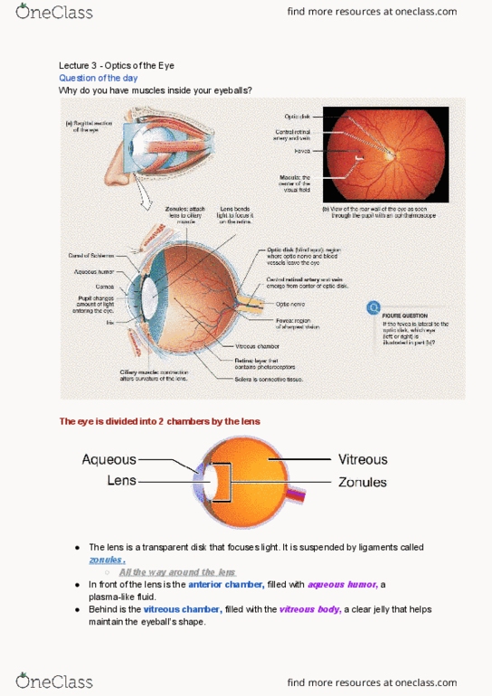
• The eye is divided into 2 chambers by the lens – the lends is a
transparent disk that focuses light. It is suspended by ligaments called
zonules
o The front of the lens is the anterior chamber, filled with
aqueous humor, a plasma-like fluid
▪ Carries nutrients and nourishes the lens
o Behind the lends is the vitreous chamber, filter with the
vitreous body, a clear jelly that helps maintain the eyeball’s
shape
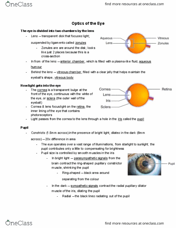
• Light enters the eyes through the cornea
o The cornea is a transparent bulge at the front of the eye,
continuous with the white of the eye, or sclera – the outer wall
of the eyeball
o The cornea and lens focus light on the retina, the inner lining of the eye that contains the photoreceptors
o Light passes from the cornea to the lens through a hole in the iris called the pupil
Pupil
• In bright light the pupils constrict (shrink) to 1.5 mm across – reducing the amount of light reaching the lens
• In the dark they dilate (enlarge) to 8 mm (~20X bigger in area) – increase the amount of light reaching the lens
• But the eye operates over a vast range of illumination, from starlight to sunlight, so the pupil contributes only a
little to compensating for brightness
• The pupil size is controlled by smooth muscles in the iris
o In bright light, parasympathetic signals from the brain contract the ring-shaped pupillary constrictor
muscles, shrinking the pupil
o In the dark, sympathetic signals contract the radial pupillary dilator muscle of the iris, dilating the pupil
• The pupil helps to focus light – like a pinhole camera
o Small pupil ensures that each point on the retina receives light from one direction in space → objects are
in focus (not blurry)
• The pupil also controls the depth of field
o When the pupil is tightly constricted, we have full depth of
field
▪ Everything we see is equally in focus
o When the pupil is dilated, we have a shallow depth of field
▪ Only objects near one specific distance are in
focus
• With pinhole-focusing, the retinal image is dim because the pinole doesn’t admit much light
o Enlarging the hole makes the image brighter but blurrier (because more light from different directions is
being admitted in)
o Using refraction helps to make the image brighter and less blurry
Refraction
• Light bends when it enters a medium with a different refractive index
• Out corneas are made of clear collagen – they bend light strongly because
there is a big different between the refractive indices of air and collagen
o The bending of light is called refraction
• In water the refraction is much weaker because the refractive indices of
collagen and water are similar
• Light is refracted by both the cornea and the lens
o Cornea is responsible for 2/3 of the eyes refraction, and the lens for 1/3
▪ Lens has the ability to change shape to adjust the focus
• The lens is a mesh of cells without nuclei, packed with clear proteins called crystallins, and are “zippered”
together in concentric layers for flexibility
find more resources at oneclass.com
find more resources at oneclass.com
Document Summary
Lecture 10: optics of the eye: the eye is divided into 2 chambers by the lens the lends is a transparent disk that focuses light. In bright light the pupils constrict (shrink) to 1. 5 mm across reducing the amount of light reaching the lens. In bright light, parasympathetic signals from the brain contract the ring-shaped pupillary constrictor muscles, shrinking the pupil. It has no blood supply relies on the aqueous humor for nutrients: the lens of the eye is convex fatter in the middle and thinner on the edges. If light hits a lens at a right angle there will be no bending: changing the shape of a lens can change the angle of incidence and change how much the light bends. Accommodation: a rounder lens bends light more and has a closer focal point, for clear vision, the focal point must fall on the retina.