Anatomy and Cell Biology 3309 Lecture Notes - Lecture 10: Calcification, Fetus, Dermal Bone

28 Dec 2017
School
Department
Professor
Document Summary
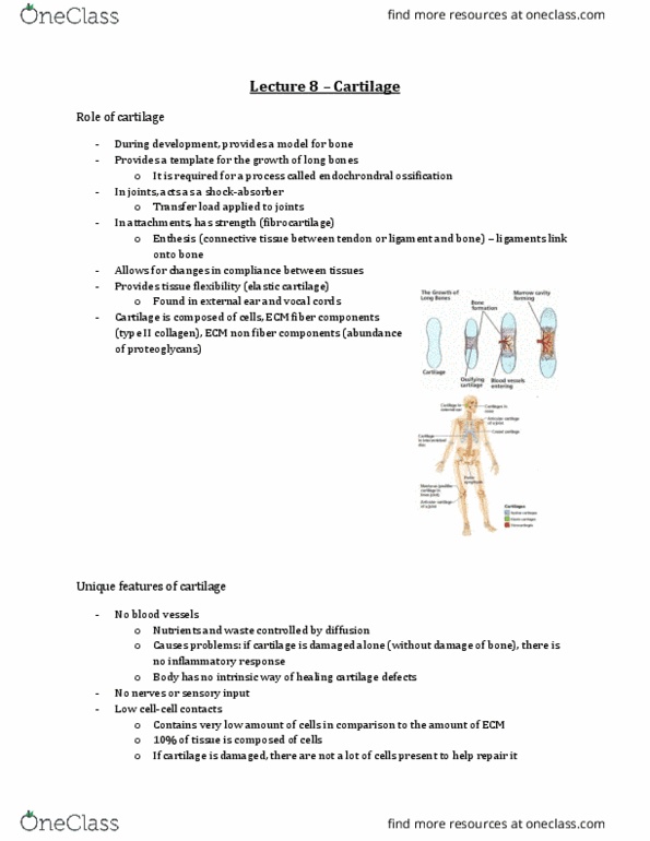
There are two methods to which bone forms in the body. Intramembranous ossification: growth of skull, maxilla and most of mandible: within mesenchyme. Endochondral ossification: growth of long bones, vertebrae, pelvis and base of skull: within cartilage model, bone formation that requires a cartilage template, area where the bone is going to form is initially cartilage. Image: bone has begun the process of endochondral ossification, top part is cartilage, lower part in diaphysis is already turning into bone. In humans, the process of endochondral ossification starts in week 6-7 of life. There are two types of growth of hyaline cartilage. Interstitial growth: mitosis of chondrocytes within cartilage unit, synthesis of new cartilage matrix. Appositional growth: differentiation of cells within the perichondrium to chondrocytes, differentiation on the outside of the chondroblasts which become chondrocytes, deposition of new cartilage matrix upon the existing cartilage. Perichondrium is on the outside where the chondroblasts are.