Anatomy and Cell Biology 3309 Lecture Notes - Lecture 40: Retinal Pigment Epithelium, Neural Tube, Ciliary Body

2 May 2018
School
Department
Professor

The Eye
Objectives
1. Understand the organization of the eye as an organ
2. Understand the development of the eye derived from the three different germ layers
3. Understand the tissue organization of the cornea, lens, tunics, iris and ciliary body
4. Relate cell specialization to function
5. Track the fate of light that enters the eye
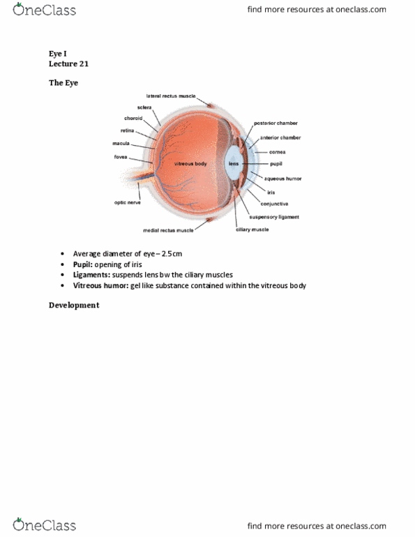
The Eye
- Diameter of the human eye = 2.5 cm
- Tunic layers
o Outer layer = sclera
o Choroid
o Retina = inner most layer
- When we talk about structures in the eye;
o Inner – relates toward the centre of the eye
o ANYTHING THAT IS TWD CENTRE OF EYE = INNER
o ANYTHING THAT IS TWD OUTSIDE OF EYE = OUTER
- In the animal kingdom, the eyes look structurally similar BUT they develop differently
o Ex. octopus eye looks structurally same as mammalian eye – ONLY LOOKS LIKE IT IS MADE
THE SAME
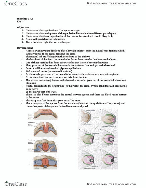
Development
- As the nervous system develops, if you have the embryo, there is a neural tube forming, which gives
rise to the spinal cord and the brain
o The neural tube is folding from the ectoderm of the embryo (in fold)
o Neural tube forms and detaches from the layer of the embryo
- Neural tube becomes the spinal cord
o At the cranial end of the fetus, it becomes the brain
find more resources at oneclass.com
find more resources at oneclass.com

- Head end of the fetus (neural tube) forms vesicles that become the brain
o One of the vesicles then forms other vesicles, that will become the retina
o Grow out of the neural tube, toward the surface of the embryo (at the head end)
- Vesicles grows OUT of the neural tube toward the surface
o It starts to invaginate & at the same time, the outer surface begins to form the lens (also
invaginates)
o The ectoderm (outer layer) will eventually become the lens
o What grew out of the neural tube, the formerly round bubble becomes like a cup
- TWO LAYER
o They lie on top of one another
o Green = retinal pigment epithelium
o Pink= neural retina
▪ Use for vision
- It is still connected to the neural tube (rest of the brain), by the optic nerve
o Optic nerve is directly connected to the rest of the brain
- CNS: retina, retinal pigment epithelium, optic nerve!!
o Part of the brain that grew OUT of the brain
- There is a blood- retina barrier
- Parts of the eye from the real ectoderm: lens, epithelium of the cornea
- Other parts of the eye are derived from mesenchymal structures
The tunics of the eye
- SCLERA = MOST OUTER LAYER
o Outer relative to the center of the eye
o Tough layer that surrounds the eye
o Keeps the eye in shape, protects it
o Continuous with the dura mater of the brain
o Continuous with the cornea at the front of the eye
- UVEA
o Contains the choroid (layer that contains blood vessels), cilia body, iris
o Vascular layer!
o Similar structure to choroid arachnoid of the brain
find more resources at oneclass.com
find more resources at oneclass.com

- RETINA = MOST INNER TUNIC
o Layer grows out of the brain, forms the optic nerve and retina
o Contains retinal pigment epithelium and neural retina (part of the eye that we use to see)
▪ TWO LAYERS – has folds
o Continuous as the double layer of epithelial cells at the inner edge of the ciliary body and the
iris
▪ THEY ARE PART OF THE RETINA
o Derive from the CNS structure
- Corneoscleral coat (cornea and sclera, ~1mm thick, protects and shapes the eye)
- Uvea (choroid, ciliary body and iris, vascular layer)
- Retina (Retinal Pigment Epithelium (RPE) and neural retina)
The Choroid
- IMAGE
o Shows the entire tunic of the eye
- Choroid is separated from the retina by Bruch’s membrane
- Bruch’s membrane
o Inner most
o Basal lamina of opposing retinal pigment epithelium and endothelial cells
o Separates the retinal pigment epithelium from the choroid
- Then you have the choroid
- Choroid contains two different layers
o Choriocapillaris – more inner portion
o Choroidal stroma – more outer layer
- Choriocapillaris
o Fenestrated capillaries
o Supplies nutrients to the outer retina
▪ Important to supply the nutrients to the outer part of the retina, which is not
vascularized in humans (No blood vessels in the outer retina)
▪ All the nutrients have to be supplied via diffusion from the choroid
- Choroidal stroma
o Contains large vessels, nerves, collagen, fibroblasts and melanocytes
- BOTH layers of choroid contain melanocytes
o They are VERY pigmented
find more resources at oneclass.com
find more resources at oneclass.com
Document Summary
Diameter of the human eye = 2. 5 cm. Tunic layers: outer layer = sclera, choroid, retina = inner most layer. When we talk about structures in the eye: anything that is twd centre of eye = inner, anything that is twd outside of eye = outer. Inner relates toward the centre of the eye: ex. octopus eye looks structurally same as mammalian eye only looks like it is made. In the animal kingdom, the eyes look structurally similar but they develop differently. Neural tube becomes the spinal cord: at the cranial end of the fetus, it becomes the brain. Vesicles grows out of the neural tube toward the surface. Two layer: they lie on top of one another, green = retinal pigment epithelium, pink= neural retina, use for vision. It is still connected to the neural tube (rest of the brain), by the optic nerve: optic nerve is directly connected to the rest of the brain.