Anatomy and Cell Biology 3309 Lecture Notes - Lecture 20: Common Bile Duct, Bile Canaliculus, Common Hepatic Duct

22 May 2018
School
Department
Professor

Histology 3309
Gall Bladder and Exocrine Pancreas
Learning Objectives
1. Describe the histology and the function of the gall bladder
2. State the function of the exocrine pancreas
3. Describe the structure of the pancreatic acinus
4. List some digestive enzymes produced by the pancreas
5. Name two duodenal hormones and explain how they control pancreatic activity
6. Describe the location and function of enterokinases
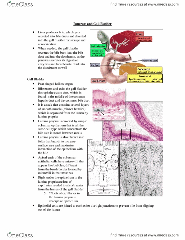
- The gallbladder is an outgrowth or an extension of the bile duct which is divided into 2 parts
o From the liver, bile is collected into the common hepatic duct
o Then it flows into the duodenum, which is now called the common bile duct
- Bile from the common bile duct reaches the duodenum at a place called the hepatopancreatic
ampulla (aka ampulla of vater)
o This duct merges with the main pancreatic duct that delivers digestive enzymes form
the pancreas
- So both bile and digestive enzymes are delivered at the same location
- The pancreas produces digestive enzymes but it also produces a bicarbonate rich fluid to
provide buffering so that enzymatic digestion with the duodenum happens at an appropriate pH
- The bile from the hepatocytes (original bile producing cells) first goes through these spaces that
represent the apical end of the hepatocytes called bile canaliculi
- The bile canaliculi are not lined by its own set of cells, it basically formed by the basal end of the
hepatocytes
- Towards the periphery of the lobule, some of the cells surrounding the bile canaliculi
differentiate into duct cells
- These channels are called bile ductules – they collect the bile towards the periphery where it
gathers more connective tissue surrounding it and into the bile duct, which is part of the portal
triad (with the portal venule and hepatic arteriole)
- YOU DON’T NEED TO KNOW THE NAME OF ALL THOSE DUCTS IN THE PIC ON THE RIGHT but
know the locatin of the gallbladder being sandwhiched bw the passageway
find more resources at oneclass.com
find more resources at oneclass.com

Gall Bladder
- Stores and concentrates bile bw meals
- At low magnification of the gallbladder, you will see that the epithelium is simple columnar
(highly columnar – the cells are very tall) that sits over a highly vascularized lamina propria of
loose connective tissue
- So you find a lot of capillaries right underneath the epithelium
- The epithelium sits on the mucosa – in the gall bladder, the mucosa forms these folds sticking
into the lumen
o These are folds and not fingerlike projections
o The way you would know that these are folds and not fingerlike projections bc we don’t
see islands
o If it was fingerlike projections, there would be islands
- Underneath the mucosa we have a smooth muscle coat
o This smooth muscle is important when delivering the bile back out into the common bile
duct after it has been stored and concentrated
- SO YOU RECOGNIZE THE GALL BLADDER WITH SIMPLE COLUMNAR EPITHELIUM FOLDS
- The gall bladder epithelium pumps Na+ and H2O
- At a low mag EM, we can see the entire epithelium
- The image on the left of the image on the right, is an inactive gall bladder
- The image on the right of the image on the right is an active gall bladder
o Spaces are appearing in bw adjacent cells
o This is due to the fact that the gall bladder epithelium has a lot of microvilli (they are not
as tall as what you will in enterocytes)
o These cells pumps Na+ out of the lumen of the gall bladder down towards the interstitial
space
o So there are a lot of Na+ pumps which actively pump Na+
o So there is an increase in Na+ concentration bw the cells and underneath the cells in the
connective tissue will result in the passive flow of water to equilibrate the osmolarity of
the high Na+ concentration
o SO water will be removed out of the lumen of the gallbladder and bile is being
concentrated
o So the bile salts will remain in the gallbladder but the water is being removed
find more resources at oneclass.com
find more resources at oneclass.com

Pancreas
- Bile enters at the hepatopancreatic ampulla together with
substances coming from the pancreas
- There is one main pancreactic duct
o Arranged like a fish bone with little ducts around it
that open into the main duct
o This arrangement means you don’t see a lot of ducts in
histological sections
- Compound acinar gland
- Acinar cells:
o Digestive enzymes
- Duct cells:
o Bicarbonate rich fluid
- Circular structure in the middle: duodenum
- The large dark stained mass of tissue on the side of it is the endocrine pancreas (you know this
bc there are little light staining dots)
- the dark staining regions make up the exocrine pancreas
o made of grape like arrangement of cells called acini
o these acini are serous secreting acini (protein producing)
o bc they produce a lot of protein, they stain dark
- the lighter areas make up the endocrine pancreas
o they are randomly organized – they don’t have a specific shape
o called the islets of langerhans
find more resources at oneclass.com
find more resources at oneclass.com
Document Summary
Bile from the common bile duct reaches the duodenum at a place called the hepatopancreatic ampulla (aka ampulla of vater: this duct merges with the main pancreatic duct that delivers digestive enzymes form the pancreas. So both bile and digestive enzymes are delivered at the same location. The pancreas produces digestive enzymes but it also produces a bicarbonate rich fluid to provide buffering so that enzymatic digestion with the duodenum happens at an appropriate ph. The bile from the hepatocytes (original bile producing cells) first goes through these spaces that represent the apical end of the hepatocytes called bile canaliculi. The bile canaliculi are not lined by its own set of cells, it basically formed by the basal end of the hepatocytes. Towards the periphery of the lobule, some of the cells surrounding the bile canaliculi differentiate into duct cells. These channels are called bile ductules they collect the bile towards the periphery where it.