Anatomy and Cell Biology 3309 Study Guide - Final Guide: Muscular Layer, Lobules Of Liver, Portal Vein

22 May 2018
School
Department
Professor


Histology 3309
Lab 17
Digestive System III
Identify the layer indicated by the brackets
- Big black bracket = Muscularis externa
- S= submucosa
o Identify with all of the glands
o Find vessels, veins, glands etc
- Dark bolded bracker = mucosa (M)
o Made of:
▪ epithelium
▪ Lamina propria
▪ Muscularis mucosa
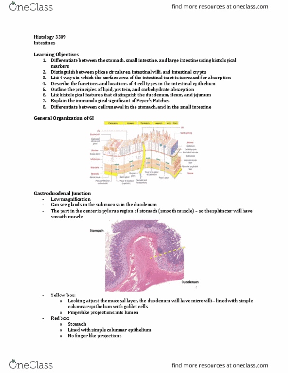
What tissue is this a section of?
- Duodenum
- Identify w brnners glands
o DON’T PUT SMALL INTESTINE – ITS DUODENUM
Identify the cell(s) indicated by the arrow
- Paneth cells
- Very very very eosinophilic
- There will be a few of these but not a ton
find more resources at oneclass.com
find more resources at oneclass.com

Identify the structure outlined by the yellow circle
- Peyers patch
- This is a section of the ileum
- Note: jejunum is lack of peyers patch and brunners gland
Identify the structure outlined by the black box
- Black bracket(M): muscularis externa
- Black box: aubachs plexus
o Bw the muscularis externa
- Meissners plexus is where the black squiggly is
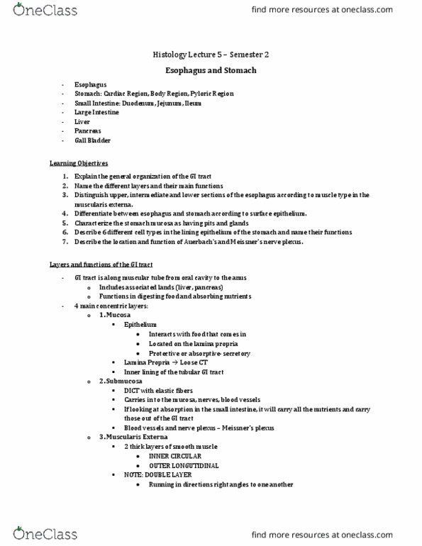
Today’s Goals
1. Identify components of portal area, the central vein and sinusoids.
2. Identify hepatocytes, Kupffer cells and endothelial cells.
3. Describe the pathway of blood throughout the liver.
4. Describe the pathway of bile from production to storage.
5. Identify the gall bladder.
6. Describe the layers of the gall bladder.
7. Identify the exocrine pancreas.
8. Identify pancreatic acinar (secretory) cells, centro- acinar cells and duct cells.
9. Explain the staining pattern of the secretory cells in terms of cellular ultrastructure.
Liver
- This is low mag (15-20x)
- See a liver lobule (outlined in black)
o Hexagonal/pentagonal in shape
o Each liver lobule will be about the same size (pretty uniform tissue)
- At the corner each hexagon/pentagon, you will find the portal triad
find more resources at oneclass.com
find more resources at oneclass.com

- Central vein in center of lobule
- This is the very center of the liver lobule
- White is the lumen of the central vein (CV)
o The central vein is lined by endothelial cells (green arrow)
o The nuclei of endothelial cells bulge into the lumen
- Coming off the central vein, we have hepatocytes
o Thee hepatocytes radiate off the central vein outward (yellow)
o They are arranged in chords
- In bw the hepatocytes, you may see light spaces →these are sinusoids (yellow arrows)
- This moth eaten appearance seen within the hepatocytes is caused by glycogen (yellow circular
outline)
- Nuclei of hepatocytes are going to have some euchromatin and heterochromatin
- This is stained with PAS (loves glycogen)
- So in this pic, instead of the glycogen not being stained and being white the glycogen in this pic
is dark staining
find more resources at oneclass.com
find more resources at oneclass.com
Document Summary
Identify with all of the glands: find vessels, veins, glands etc. Dark bolded bracker = mucosa (m: made of, epithelium, lamina propria, muscularis mucosa. Identify w brnners glands: don"t put small intestine its duodenum. There will be a few of these but not a ton. Identify the structure outlined by the yellow circle. This is a section of the ileum. Note: jejunum is lack of peyers patch and brunners gland. Identify the structure outlined by the black box. Black box: aubachs plexus: bw the muscularis externa. Meissners plexus is where the black squiggly is. Identify components of portal area, the central vein and sinusoids. Identify pancreatic acinar (secretory) cells, centro- acinar cells and duct cells. See a liver lobule (outlined in black: hexagonal/pentagonal in shape, each liver lobule will be about the same size (pretty uniform tissue) At the corner each hexagon/pentagon, you will find the portal triad. This is the very center of the liver lobule.