Anatomy and Cell Biology 3309 Lecture Notes - Lecture 10: Hyaline Cartilage, Bronchus, Trachea

29 Apr 2021
School
Department
Professor
Document Summary
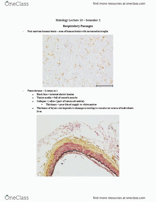
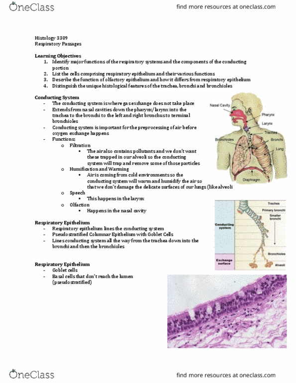
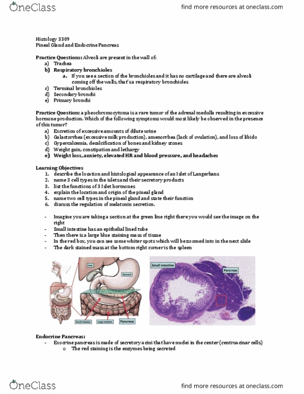
In the trachea and extrapulmonary bronchi the hyaline cartilage is organized into rings. In the intrapulmonary bronchi the hyaline cartilage is organized into anatomizing plates. This is an image of the intrapulmonary bronchus - you can tell by the alveoli at the bottom. There is smooth muscle under the epithelium too - this is not the trachealis muscle. Histological features of the bronchial tree: summary recap. Can see that within the lung the cartilage is broken up. The cartilage is also getting smaller, being replaced by smooth muscle and elastic fibres. At one point when the air pipe gets smaller, the epithelium changes to simple columnar and then to simple cuboidal. The cartilage is the best way to determine if we are looking at bronchus or bronchiole. Once we get into the respiratory part, the ciliated cells disappear and smooth muscle eventually too.