Foods and Nutrition 1021 Lecture Notes - Lecture 17: Osteoma, Splints, Gadolinium

9 Dec 2017
School
Department
Course
Professor
Document Summary
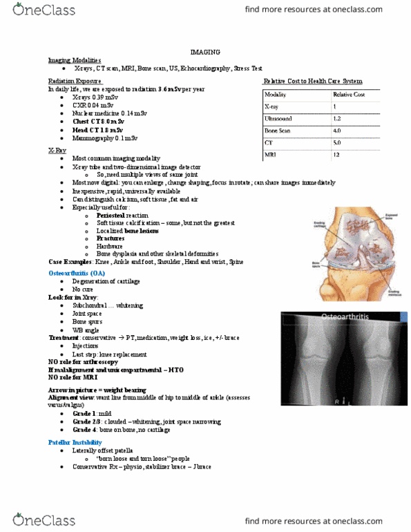
Imaging modalities: x-ray, ct, mri, bone scan, ultrasound, echocardiography, stress test. In daily life, we are exposed to 3. 6msv per year: xray = 0. 39msv, cxr = 0. 04msv, nuclear medicine = 0. 14msv, chest ct = 8. 0msv, head ct = 1. 8msv, mammography = 0. 1msv. X-ray tube and two dimensional image detector we need multiple views of the same jt because it is only 2d. Most are now digital, can enlarge it, change shade, focus in, rotate, share results immediately through phone: inexpensive, rapid, universally available, can distinguish ca (bone, kidney stones etc. For: periosteal reaction, soft tissue calcification, localized bone lesions, fractures, hardware, bone dysplasia and other skeletal deformities. This is when you get degeneration of the cartilage in a joint, common in the knee. What we see: subcondylar sclerosis = whitening below condyles, jt. space = usually less jt. space, bone spurs, knee angle or weight bearing angle of the legs.