Kinesiology 2222A/B Lecture Notes - Lecture 23: Brachiocephalic Artery, Brachiocephalic Vein, Axillary Artery

7 Feb 2016
School
Department
Course
Professor

Last Anatomy Lecture 2016-02-08 12:07:00 AM
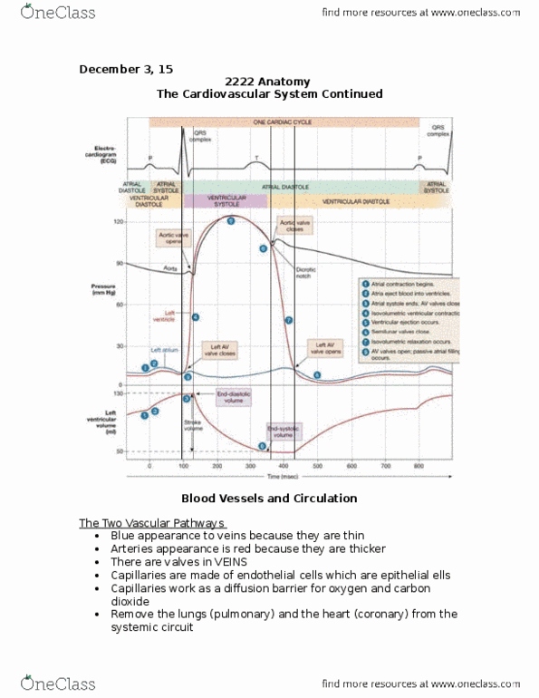
Blood Vessels and Circulation:
The Two Vascular Pathways
• Arteries
o Higher pressure
o Away from the heart
• Veins
o Characteristically blue
o Arteries are thin, this gives them a blue colour
o Blood to the heart
o Inside veins we have valves → important to ensure one
directional flow
• Capillaries:
o Connect arteries and veins
o Where gas exchange occurs → oxygen is delivered from the
tissues and CO2 is removed from the veins
o Made up on endothelial cells → single cells that work as a
diffusion barrier
Systemic Arteries:
• The systemic circuit does NOT involve the lungs
• Provide blood to everything but the heart itself and the lungs
Ascending Aorta – Aorta Arch Review:
• Aorta turns into the aortic arch
• The first branch is the brachiocephalic trunk
o we only have 1
o On the right side of the vertebral column
o Extends over the vertebrae to make it symmetrical
o We run into the left common carotid and the left Subclavian
that come off of the Brachiocephalic trunk
▪ These vessels provide blood to the head, neck, upper
trunk and upper limbs → most of the body receives
blood from this area
Head and Neck Arteries:
• When a vessel branches we sometimes change the name of it
• Common = more branches to come
find more resources at oneclass.com
find more resources at oneclass.com

• Off of the common carotid there is the external carotid and internal
carotid
• Off of the right Subclavian you have a small artery that comes off of
it called the vertebral artery that goes through the transverse
process of the cervical vertebrae
Brain Arteries:
• Circle of Willis sits at the bottom of the brain and it is where the
vertebral arteries feed into to provide blood to the brain
• As long as you provide blood through one of these roots there is
enough blood to deliver to the brain
• This is known as anastomosis which prevents blood loss to the brain
to ensure adequate flow
• Anastomosis is the meeting of arteries to ensure collateral blood
flow in the case of blockage
Upper Limb Arteries:
• There is basically one vessel that provides blood flow to regions
that just splits and changes name
• The Subclavian becomes the Axillary artery when it goes under the
clavicle (axillary = armpit)
• The Axillary artery becomes the Brachial artery once it touches the
humerus
• When the Brachial Artery reaches the radius and ulna it branches
into the radial artery and ulnar artery
Abdominal/Descending Aorta:
• Extending off of the aorta
• There are some branches that go through the rib cage and are
called intercostal arteries
• There are many little arties that arise off the descending aorta that
supply blood to our internal organs
• As it passes the diaphragm its name changes into the abdominal
aorta
• as it progresses down into the pelvic region it divides around L4
into a right and a left common iliac artery
3 Unpaired Abdominal Arteries:
find more resources at oneclass.com
find more resources at oneclass.com

• Unpaired = just one
• Celiac Trunk:
o First major branch off of the abdominal aorta
o Mostly responsible for the digestion process
o Provides blood to the stomach, liver, spleen, gall bladder,
pancreas and duodenum
• Mesenteric arteries are responsible for the intestines
• Superior Mesenteric:
o Feeds most of the intestines
• Inferior Mesenteric:
o Supplies the descending colon, sigmoid and rectum
3 Paired Abdominal Arteries:
• Renal Arteries:
o Below the adrenal
o 2 kidneys that each receive blood through the renal artery
• Adrenal Arteries:
o Sit on top of the kidneys
o Also known as suprarenal
o Send blood to the adrenal gland
o responsible for stress
• Gonadal Arteries:
o Covers both the testicular artery and the ovarian artery
o Originate off of descending aorta
Arterial Supply to the Thigh:
• Branches into the left and right common iliac artery
• The common iliac turns into the external and internal iliac arteries
• Internal Iliac:
o Goes inside the pelvis → internal
o Provides blood to the internal structures within the pelvis →
reproductive organs, the bladder, etc.
• External Iliac:
o carries on to feed blood to the lower limb
o The second that it passes the inguinal ligament it becomes
the femoral artery which provides blood the quads,
hamstrings, groin, etc.
find more resources at oneclass.com
find more resources at oneclass.com
Document Summary
Systemic arteries: the systemic circuit does not involve the lungs, provide blood to everything but the heart itself and the lungs. Systemic artery: the vein beside the artery will have the same name, they don"t have the same directional flow of the heart. Venous circuit: often travel with arteries and have corresponding names, differences, return of blood to the heart, superficial veins, blood in the brain, collected by dural sinuses, blood in the abdomen (hepatic portal system) Head and neck veins: similar external and internal vessel taking blood from outside of the skull and the inside of the skull, external jugular feeds into the brachiocephalic trunk, internal jugular feeds into the brachiocephalic vein. Dural sinuses: collect blood from the brain that has been used and returns it to the internal jugular vein, many entry points, feeds into the internal jugular vein which feeds into the brachiocephalic brain.