Kinesiology 3336A/B Lecture Notes - Lecture 15: Subtalar Joint, Anatomical Terms Of Location, Shin Splints

19 Mar 2017
School
Department
Course
Professor
Document Summary
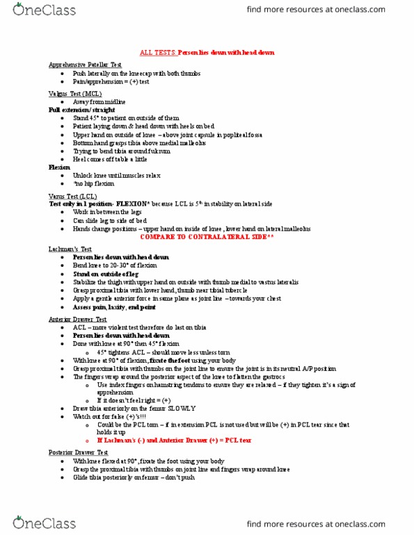
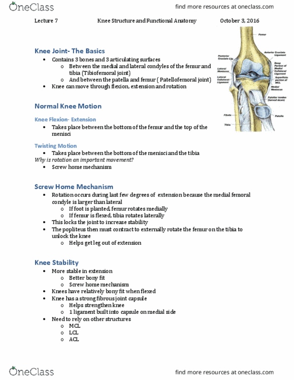
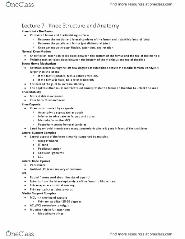
Knee surface anatomy: medial and lateral femoral condyles, head of fibula, tibial tuberosity, anterior joint line, gerdy"s tubercle, mcl, lcl attaches to fibular head, medial and lateral meniscus, biceps femoris tendon. It band first attaches to gerdy"s tubercle. Lcl + biceps femoris attach to head of fibula. Apprehensive patellar test: push laterally on the kneecap with both thumbs, pain/apprehension = (+) test. Valgus test (mcl: bring leg away from midline. Flexion: unlock knee until muscles relax, *no hip flexion. If lachman"s (-) and anterior drawer (+) = pcl tear. Posterior drawer test: with knee flexed at 90 , fixate the foot using your body, grasp the proximal tibia with thumbs on joint line and fingers wrap around knee, glide tibia posteriorly on femur don"t push. If medial meniscus external rotation of foot, knee extends in opposite way of heel (varus) If lateral meniscus internal rotation of foot, knee extends in opposite way of heel (valgus)