Kinesiology 3337A/B Lecture Notes - Lecture 10: Atrioventricular Node, Luigi Galvani, Purkinje Fibers

22 Oct 2016
School
Department
Course
Professor
Document Summary
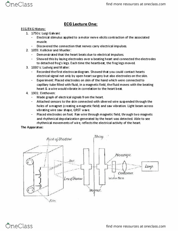
Light beam across vibrating wire saw shape (1901) qrst wave. Tracings reflect voltage changes in the body (depolarization) Vibrations in magnetic field projected to see movement of wire as electro activity moved through. Myocardium- no origin and insertion on bones- myoctes attach to adjacent fibers. The myocardial cells function together to produce a coordinated contraction of the entire organ. Myocardial cells will depolarize- move through the heart quickly. Myocardial cells are connected to each other by gap junctions. These allow action potentials to spread quickly: depolarization wave hopes from one myocyte to the next- enabling heart to contract synchronously. Specific arteries supply blood to sa node. The excitation from the sa node (conduction system cells with the fastest rate of spontaneous depolarization- pacemaker cells) Sa node- surrounds the nodal artery from which it receives a rich blood supply. These smaller, modified muscle cells generate the electrical signal that controls the heart.