Physiology 2130 Lecture Notes - Lecture 31: Resting Potential, Atrioventricular Node, Purkinje Fibers

1 May 2018
School
Department
Course
Professor

Lecture 031: Excitation and electrical activity of the cells
Objectives
● Origin of self-excitability:
● APs can spontaneously generated in the SA node (pacemaker)
● Conducting system of the heart
● ECG
● Ventricular muscle APs
Myocardial Cells
● Contractile cells (atrial and ventricular muscles)
○ Short and branched
○ Contain GAP junctions
■ Allows APs to jump from one cell to the nex
■ Very important in conduction of the AP
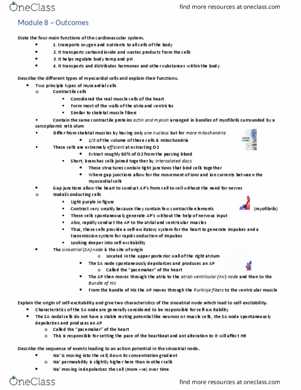
● Specialized excitatory (nodal) and conducting cells
○ Excitatory nodal cells
■ SA node
■ AV node
○ Conducting cells
■ Bundles of His
■ Purkinje fibers
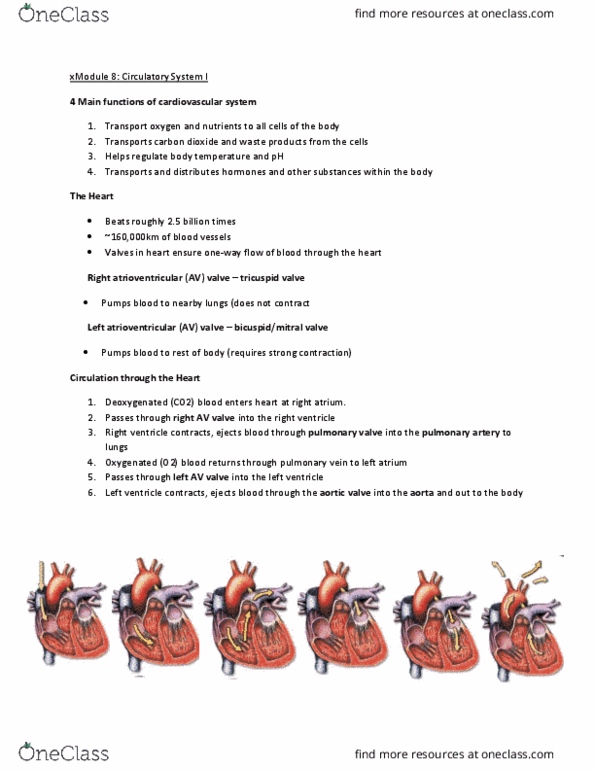
The origin of self-excitability
● Normal heart rate (at rest) is 72 bpm (in vivo)
● Impulses (normally) originate in the sinoatrial (SA) node located in the upper posterior
wall of the right atrium
● Most cells in the heart have the capability to self-excite
○ Can spontaneously produce APs
● However, the SA node has the greatest/fastest rate of self-excitation
○ Once it is generated the AP “sweeps” through the rest of the heart and override
the cells that are slowly being to generate excitation
● Thus the SA node is the “PACEMAKER” of the heart
● There are many types of APs in the heart
○ Different cardiac cells use special ion channels to produce distinctive APs
■ Or the same type of channels in a different way/time
■ Level of depolarization depends on the type of AP produced
○ SA node
■ Slow response AP
■ The depolarizing phase is slower than the fast-response AP
○ Ventricular muscle
■ Fast response AP
○ Note: AP firing rates are slower in the heart (measured in seconds rather than
milliseconds)
find more resources at oneclass.com
find more resources at oneclass.com

Chemical composition Inside and outside a cell
● Inside: K+
● Outside: Na+, Ca2+, Cl-
SA node-characteristic responsible for self-excitation
● Heart is different than the nerve or muscle cell in this
aspect
● These cells have a much GREATER Na+ and Ca2+
permeability
○ In other words, there is a slow positive inward current when the heart is in
diastole
● In addition, there is also a DECREASE IN K+ during diastole (relaxation)
○ Contributes to a slow depolarization towards threshold
■ Slowly spontaneously depolarize towards threshold
● SA nodal cell do not have a stable “resting” membrane potential
○ Have a PRE-POTENTIAL or PACEMAKER POTENTIAL
■ Membrane potential varies between -60 mV to +20 mV
● due to the slow depolarization
■ Has a threshold voltage of -40 mV
● In diastole (rest)
○ Pre-potential
■ A slow spontaneous depolarization caused by a slow build-up of
positive charges inside the cell due to 3 things
● SPONTANEOUS
1. The increase of Na+ permeability into the cell
● through Na+ funny channels
○ These voltage-gated channels are “funny” because they
open when the membrane potential returns to -60 mV
(when membrane repolarizes)
2. The increase of Cal2+ permeability into the cell
● Through T-type voltage-gated channels (most)
○ T: Transient (very brief)
● And slow L-type voltage-gated channels (some)
○ L: long-lasting
3. The decrease of K+ permeability out of the cell
● Outward K+ movement decrease over time
○ Depolarizing phase
■ At threshold (-40 mV)
● Na+ funny channels and T-type Ca2+ voltage-gated channels
CLOSE
● ALL L-type Ca2+ voltage-gated channels OPEN
■ Ca2+ flows in
● Does so “slowly”, thus SA are slow response AP
find more resources at oneclass.com
find more resources at oneclass.com
Document Summary
Lecture 031: excitation and electrical activity of the cells. Aps can spontaneously generated in the sa node (pacemaker) Allows aps to jump from one cell to the nex. Very important in conduction of the ap. Normal heart rate (at rest) is 72 bpm (in vivo) Impulses (normally) originate in the sinoatrial (sa) node located in the upper posterior wall of the right atrium. Most cells in the heart have the capability to self-excite. However, the sa node has the greatest/fastest rate of self-excitation. Once it is generated the ap sweeps through the rest of the heart and override the cells that are slowly being to generate excitation. Thus the sa node is the pacemaker of the heart. There are many types of aps in the heart. Different cardiac cells use special ion channels to produce distinctive aps. Or the same type of channels in a different way/time. Level of depolarization depends on the type of ap produced.