Physiology 2130 Lecture Notes - Lecture 53: Respiratory Center, Cerebral Cortex, The Automatic

1 May 2018
School
Department
Course
Professor

Lecture 053: Regulation of respiration
True or False
● The pressure-volume relationship of the lung in humans can only be measured by the
helium dilution method
○ FALSE
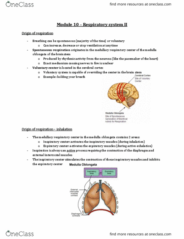
Control of respiration: the voluntary system
● Change how you breath
● Signals from cerebral cortex -> corticospinal tract -> spinal neurons -> respiratory
muscles
● This is a limited process
○ Ultimately, the automatic system will take over
○ You can only hold your breath for so long
The Automatic System
● Maintains normal blood gas values under a variety of condition
○ Very efficient control
● Receptors
○ Gather information about the current state of blood gases and other factors
(negative feedback)
○ Ends input signals to the central controller
● Central controller
○ Coordinates and responds to the information provided by the receptors
○ Generates a response to send to the respiration muscles to get a desired result
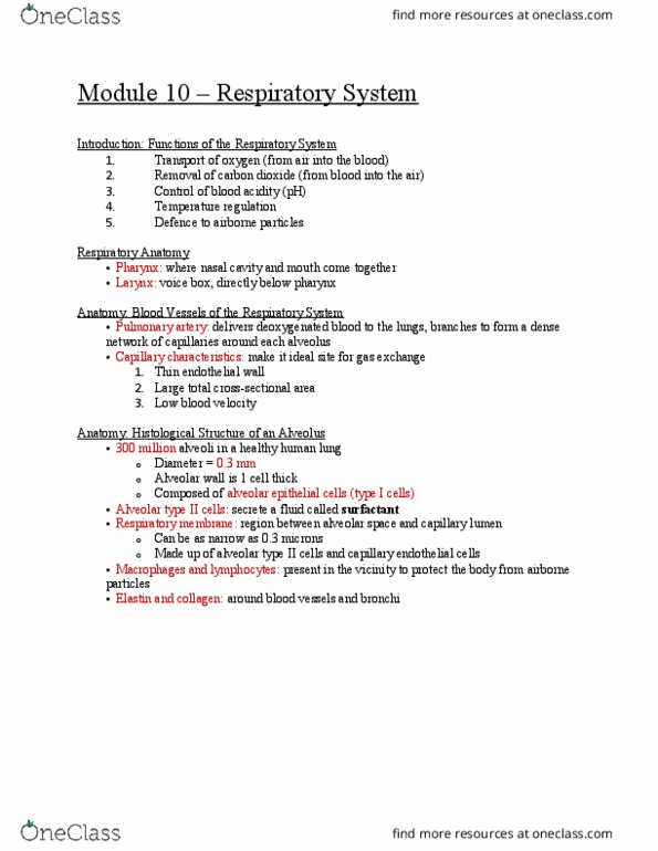
● Respiratory muscles (diaphragm and intercostals)
○ Receive impulses from the controller to ultimately affect ventilation
The central controller
● Neurons located in 4 areas
○ Inspiratory area
○ Pneumotaxic area
○ Apneustic center
○ Expiratory area
● Lesions to these areas results in respiratory pathologies
○ How we know these areas are involved in normal respiration
1. Inspiratory area
● Location:
○ Dorsal group of neurons in the medulla that send signals to muscles to initiate
inspiratory drive
● Properties:
○ Critical in respiration !!
○ Inherent rhythmic excitability
find more resources at oneclass.com
find more resources at oneclass.com

○ Initiates inspiratory drive
2. Pneumotaxic area
● Location:
○ Group of neurons in the pons
● Properties:
○ Fine-tuning of respiration
■ Not essential
○ Limits duration of inspiratory drive “Switch off”
○ Control of inspiratory volume and respiratory rate
○ Without this center you will be gasping for air instead of taking subtle breaths
3. Apneustic center
● Location:
○ Group of neurons in the lower pons
● Properties:
○ Not sure what it exactly does...
○ Opposite of “pneumotaxic center”
■ Can prolong inspiration
● Cause long, gasping breaths
■ However, the pneumotaxic center overrides apneustic center
4. Expiratory area
● Location:
○ Ventral group of neurons in the medulla
● Properties:
○ When needed (remember expiration is normally a passive process), these
neurons send signals to expiratory muscles resulting in active expiration
find more resources at oneclass.com
find more resources at oneclass.com
Document Summary
The pressure-volume relationship of the lung in humans can only be measured by the helium dilution method. Signals from cerebral cortex -> corticospinal tract -> spinal neurons -> respiratory muscles. Ultimately, the automatic system will take over. You can only hold your breath for so long. Maintains normal blood gas values under a variety of condition. Gather information about the current state of blood gases and other factors (negative feedback) Ends input signals to the central controller. Coordinates and responds to the information provided by the receptors. Generates a response to send to the respiration muscles to get a desired result. Receive impulses from the controller to ultimately affect ventilation. Lesions to these areas results in respiratory pathologies. How we know these areas are involved in normal respiration: inspiratory area. Dorsal group of neurons in the medulla that send signals to muscles to initiate inspiratory drive. Limits duration of inspiratory drive switch off .