Physiology 2130 Lecture Notes - Lecture 8: Sympathetic Nervous System, T Wave, Parasympathetic Nervous System

5 Jul 2018
School
Department
Course
Professor

Module 8 – Circulation Part 1: The Heart
Intro:
Some fun facts
oThe heart is one of the most remarkable organs in the body
oThe heart is about the size of your fist
oThe heart sits in our chest cavity between your lungs
oDuring the course of an average life the heart will beat 2.5 billion times
oYou have roughly 160 000 km of blood vessels that transport blood to almost every cell in your body
Four principle functions of the cardiovascular system
o1. transports oxygen and nutrients to all cells of the body
o2. It transports carbon dioxide and wastes products from the cells
o3. It helps regulate body temp and pH
o4. It transports and distributes hormones and other substances within the body
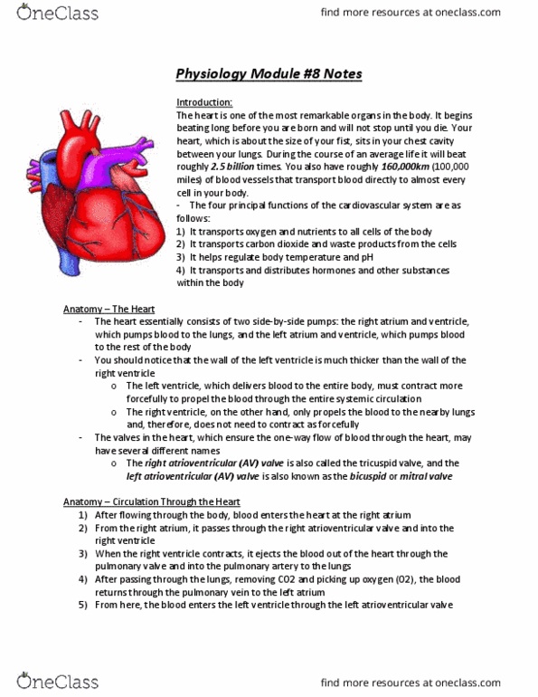
Anatomy of the Heart:
The heart consists of two side-by-side pumps
oThe right atrium and ventricle – pumps blood to the lungs
oThe left atrium and ventricle – pumps blood to the rest of the body
The walls of left ventricle is much thicker than the walls of the right ventricle
oThe left ventricle delivers blood to the entire body and must contract much more forcefully
oThe right ventricle only pumps blood to the lungs close by
The valves of the heart ensure one-way blood flow through the heart
oThe right atrioventricular (AV) valve or tricuspid valve
oThe left atrioventricular (AV) valve or bicuspid valve or mitral valve
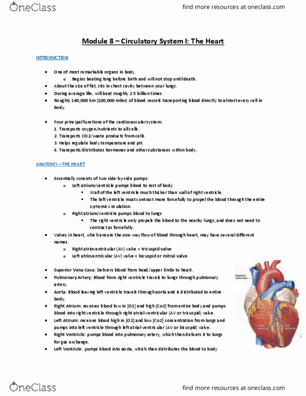
Anatomy of the Heart Detailed:
Superior vena cava: delivers blood to the heart from the head and upper limbs
Inferior vena cava: delivers blood to the heart from the lower body
Right atrium: receives blood from the entire body and pumps blood to the right ventricle through the right
AV (or tricuspid) valve
oBlood entering the right atrium is low in O2 and high in CO2
Right AV (tricuspid) valve: ensures blood flows in one direction from the right atrium to the right ventricle
Right ventricle: pumps blood into the pulmonary artery to be sent to the lungs for gas exchange
Pulmonary valve: ensures blood flows in one direction from the right ventricle to the pulmonary artery
Pulmonary artery: blood leaving the right ventricle travels to the lung through this
Left atrium: receives blood that has come from the lungs and pumps blood to the left ventricle through the
left AV (or bicuspid) valve
oThis blood is rich in O2 and low in CO2
Left AV (bicuspid) valve: ensures blood flows in one direction from the left atrium to the left ventricle
Left ventricle: pumps blood into the aorta to be sent to the entire body
Aortic valve: ensures blood flows in one direction from the left ventricle to the aorta
Aorta: blood leaving the left ventricle travels through this and is distributed to the entire body
Chordae tendineae: cords of collagen that attach to the valves at one end and to papillary muscles at the
other
oPrevents the AV valves from being pushed into the atria when ventricle pressure is high
Papillary muscles: extensions of the ventricular muscles and are attached to the chordae tendineae
oHolds the AV valves in place during contraction
Circulation Through the Heart:
Body right atrium right AV valve right ventricle pulmonary valve pulmonary artery lungs
find more resources at oneclass.com
find more resources at oneclass.com

pulmonary vein left atrium left AV valve left ventricle aortic valve aorta body
Myocardial Cells:
Two principle types of myocardial cells
oContractile cells
Considered the real muscle cells of the heart
Form most of the walls of the atria and ventricles
Similar to skeletal muscle fibers
Contain the same contractile proteins actin and myosin arranged in bundles of myofibrils surrounded by a
sarcoplasmic reticulum
Differ from skeletal muscles by having only one nucleus but far more mitochondria
1/3 of the volume of these cells is mitochondria
These cells are extremely efficient at extracting O2
Extract roughly 80% of O2 from the passing blood
Short, branches cells joined together by intercalated discs
These structures contain tight junctions that bind cells together
Where gap junctions allow for the movement of ions and ion currents between the
myocardial cells
Gap junctions allow the heart to conduct AP’s from cell to cell without the need for nerves
oNodal/conducting cells
Light purple in figure
Contract very weakly because they contain few contractile elements (myofibrils)
These cells spontaneously generate AP’s without the help of nervous input
Also, rapidly conduct the AP to the atrial and ventricular muscles
Thus, these cells provide a self-excitatory system for the heart to generate impulses and a
transmission system for rapid conduction of impulses
Looking deeper into self-excitability
The sinoatrial (SA) node is the site of origin
oLocated in the upper posterior wall of the right atrium
The SA node spontaneously depolarizes and produces an AP
oCalled the “pacemaker” of the heart
The AP then moves through the atria to the atrial-ventricular (AV) node and then to the
Bundle of His
From the bundle of His, the AP moves through the Purkinje fibers to the ventricular muscle
Note: Myocardial cells and our favourite ions
These ions – Na, Cl, Ca, and K – are also responsible for the AP in the heart
Note: The absolute cause of the spontaneous AP is still controversial
SA Node Action Potential:
Characteristics of the SA node is generally considered to be responsible for self-excitability
Na+ is moving into the cell, down its concentration gradient
oNa+ permeability is slightly higher here than in other cells
Na+ moving in depolarizes the cell (more +ve) over time
Ca+ is similar to Na+ - moving into the cell and depolarizing
Depolarization doesn’t automatically = AP
Na+ and Ca+ contribute to the spontaneous AP, but the movement of K+ is its main cause
oK+ are trying to leave the cell down its concentration gradient
oThis hyperpolarizes the cell (more -ve)
find more resources at oneclass.com
find more resources at oneclass.com

oIf you want depolarization you do not want K+ leaving the cell
oSo, the K+ permeability of the SA node decreases over time – less K+ leaks out
Also, the Na/K pump will return K+ into the cell
Because Na+ and Ca+ are flowing into the cell and K+ builds up inside, the membrane potential of the SA node
depolarizes to -60 mV -40 mV (threshold)
oThe SA nodal cells do not have a stable resting potential like neurons or muscle cells
This slow depolarization is completely spontaneous and is called the pacemaker potential
oThis is responsible for setting the pace of the heartbeat and ant alteration to it will affect HR
Once the membrane depolarizes to threshold (-40 mV) special voltage-gated Ca+ channels will open, Ca+ will rapidly
flow into the cell, depolarization will result, and an AP will begin
The Ca+ channels begin closing when voltage-gated K+ channels begin to open, letting K+ out to repolarize the cell
Once the cell has returned to its lowest value (-60 mV) the pacemaker potential will begin depolarizing the cell and
the sequence will repeat
Note: influx of Ca+ is important in contraction (seen later)
Note: don’t need to know voltage changes
General review of above because confusing:
Na+ and Ca+ move into the SA nodal cell, causing a depolarization of the membrane
oNo AP yet
K+ permeability decreases, so less K+ can leak out
This build-up of Na+, Ca+, and K+ leads to a depolarization
o-60 mV -40 mV
Voltage-gated channels now open and Ca+ will rapidly enter the cell
oAP produced
After the AP, Ca+ channels will close while K+ channels open, repolarizing the cell
Once the cell has returned to its lowest value (-60 mV) the pacemaker potential will begin depolarizing the cell and
the sequence will repeat
Myocardial Cells – The Conducting System of the Heart:
Once the AP is generated in the SA node it travels throughout the heart in a highly coordinated
manner
Pathway
oSA through atrial muscle (downward contraction occurs) AV node (must pass through this node due to
fibrous tissue separation) down the bundle of His apex of the heart through the Purkinje fibers up
through the ventricular muscles (upwards contraction occurs)
It is very important to have a well-coordinated contraction for the heart to function properly as a pump
oTherefore, conduction speed of the AP varies as it moves through the heart
Varying speeds
oNote: don’t need to know exact speeds but know generally which is faster and slower
oSA node – one of the slowest conduction speeds (0.05 m/sec)
oAtrial muscles – speeds up to ensure muscle contracts simultaneously (1 m/sec)
oSA atrial muscles – top down contraction, blood moved down into ventricles
oAV node – slows speed of conduction to ensure atria finish contracting before ventricles contract (0.03
m/sec)
oBundle of His – very rapid conduction down to the apex of the heart (1 m/sec)
oPurkinje fibers – quick conduction of the AP (5 m/sec)
oVentricle muscles – muscles contraction bottom up, pushing blood up (1 m/sec)
Electrocardiogram (ECG):
Intro:
find more resources at oneclass.com
find more resources at oneclass.com
Document Summary
Module 8 circulation part 1: the heart. Four principle functions of the cardiovascular system: 1. transports oxygen and nutrients to all cells of the body, 2. It transports carbon dioxide and wastes products from the cells: 3. It helps regulate body temp and ph: 4. It transports and distributes hormones and other substances within the body. The heart consists of two side-by-side pumps: the right atrium and ventricle pumps blood to the lungs, the left atrium and ventricle pumps blood to the rest of the body. The valves of the heart ensure one-way blood flow through the heart: the right atrioventricular (av) valve or tricuspid valve o. The left atrioventricular (av) valve or bicuspid valve or mitral valve. Superior vena cava: delivers blood to the heart from the head and upper limbs. Inferior vena cava: delivers blood to the heart from the lower body.