Physiology 3120 Lecture Notes - Lecture 36: Theca Interna, Tunica Externa, Smooth Muscle Tissue

2 May 2018
School
Department
Course
Professor

Human Physiology Lecture 36
Blood Vessel Structure
- Blood vessel structure relates to their function
Blood Vessel Structure
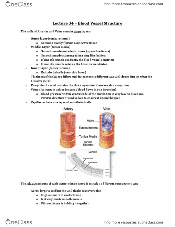
The walls of Arteries and Veins contain three layers:
- Outer layer (tunica externa)
o Fibrous connective tissue
▪ Holding things together
- Middle Layer (tunica media)
o Find specialized tissue: circularly arranged smooth muscle & elastic tissue
o Smooth muscle arranged in a ring like fashion
▪ If it contracts, it causes the blood vessel to constrict
▪ If it relaxes, it causes the blood vessel to dilate
▪ CONTRACT = CONSTRICT, RELAX = DILATE
- Inner Layer (tunica interna)
o Endothelial cells
o Thin
- Content & thickness of each of the layers are different depending on what the blood vessel is
- Almost every blood vessel contains all three layers
- Veins also contain valves
o Ensure blood flows in only one direction
o They have valves because the blood pressure in the venous side of circulation is VERY low;
there is the possibility that blood could reverse direction
- Capillaries have one layer of endothelial cells
find more resources at oneclass.com
find more resources at oneclass.com

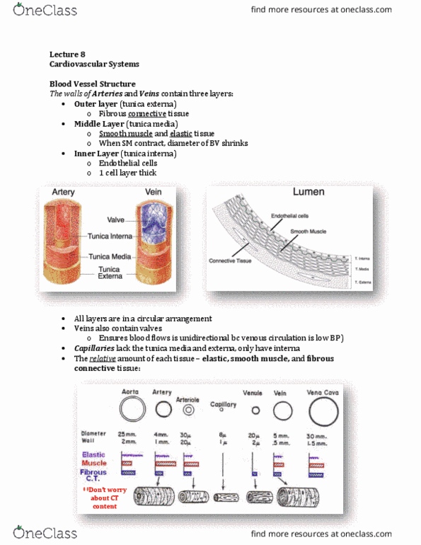
- The relative amount of each tissue: elastic, smooth muscle and fibrous connective tissue
- KNOW RELATIVE AMOUNTS & GENERAL IDEA OF RELATIVE SIZE & THICKNESS
- Aorta:
o Large diameter vessel – 25mm
o Thin wall thickness
o Walls contain A LOT of elastic tissue, some smooth muscle, and fibrous connective tissue
holding everything together
- Arteriole:
o VERY thick wall relative to their size
o Wall contains predominantly smooth muscle tissue. Smooth muscle is important for
regulating the diameter of the vessel
- Capillary:
o Smallest vessels – diameter & thin wall (one cell thickness, no outer or inner layer)
o Endothelial cell has no elastic, smooth muscle, or fibrous CT
- Vena cava:
o Larger diameter than aorta
o Wall thickness is thinner than the aorta
o Contains some smooth muscle
o Note: to increase SV, have to increase the filling of the heart, and it can be done by increasing
venous return activate SYN – activates the smooth muscle in the wall, causing the vessels
to constrict and help pump blood back to the heart & increase venous return
▪ Increase EDV to increase SV
- How does the blood vessel structure relate to its function?
Structure relates to function
- Aorta & large arteries – BP is very pulsatile
o Heart contracts, ejects blood out to the vessels
o As the vessel is getting the ejection of blood, they EXPAND
- When the ventricles contract during systole, they eject some amount of blood
o Even during rest, you eject 70mL of blood
o Blood enters the aorta and artery, since they are thin walls with a lot of elastic tissue – they
can expand to accommodate the in rush of blood
▪ VERY COMPLIANT!
find more resources at oneclass.com
find more resources at oneclass.com

- Compliance impacts pulse pressure
- Vessels can accommodate rush of blood = aorta, arteries
o Structure allows for in rush of blood, expand
o When the heart is relaxing, they may begin to contract & help continue blood along
o Since they can expand, there is VERY LITTLE resistance = small drop in BP
o Large arteries are very good at distributing blood throughout the body because of the small
BP drop= distribution vessels
o Maintain high BP throughout the vessels
- Arterioles:
o Largest drop of BP is there because it is the site of HIGHEST resistance
▪ Resistance is dependent on the size of the blood vessel & arterioles are very small
▪ Thick wall with very little elastic tissue in it – therefore it can not expand as much as
the arteries
▪ Don’t expand to accommodate in rush of blood
o Site of HIGH resistance = resistant vessels
o Have a lot of smooth muscle – allows vessels to relax, dilation or contract, constriction
o Vessels help regulate blood flow because we are altering blood flow with pressure gradient +
radius
▪ Change radius by contracting or relaxing smooth muscle of the walls
- Capillaries:
o Smallest vessels
o Short diffusion distance
o Largest total cross sectional area of ALL blood vessels
o Blood is moving at is SLOWEST = slow blood velocity
o Exchange vessels – site where gas exchange, nutrient exchange, etc.
▪ Low blood velocity, high total SA, short diffusion distance
- Veins
o Where you find most of your volume at rest – 70% of total blood volume is found in the vein
o Larger diameter than their arteriole counterparts – therefore can accommodate more blood
o Thinner walls than arterioles counterparts
o Easy to expand due to the elastic tissue content
o Capacitance/collection vessels – contain majority of the total blood volume
o Contain smooth muscle – can be controlled by ANS
▪ Can control size, constrict
o Have valves to ensure when you do squeeze them, blood can only go in one direction: back to
the heart
o Help regulate cardiac output by changing venous return
▪ Increase venous return increase EDV increase SV increase CO
CV Regulatory Mechanisms
- Purpose: homeostasis (maintain relatively constant conditions of the internal environment)
o 1. To increase blood flow to active tissues
▪ CO is matched to your oxygen demands in your body – as the demand goes up, you
have to increase blood flow/ CO
• If a tissue needs more oxygen, the tissue can regulate its own blood flow
o 2. To increase/decrease heat loss from the body
find more resources at oneclass.com
find more resources at oneclass.com
Document Summary
Blood vessel structure relates to their function. The walls of arteries and veins contain three layers: Outer layer (tunica externa: fibrous connective tissue, holding things together. Middle layer (tunica media: find specialized tissue: circularly arranged smooth muscle & elastic tissue, smooth muscle arranged in a ring like fashion. If it contracts, it causes the blood vessel to constrict. If it relaxes, it causes the blood vessel to dilate: contract = constrict, relax = dilate. Inner layer (tunica interna: endothelial cells, thin. Content & thickness of each of the layers are different depending on what the blood vessel is. Almost every blood vessel contains all three layers. Veins also contain valves: ensure blood flows in only one direction, they have valves because the blood pressure in the venous side of circulation is very low; there is the possibility that blood could reverse direction. The relative amount of each tissue: elastic, smooth muscle and fibrous connective tissue.