Physiology 3120 Lecture Notes - Lecture 5: Pulmonary Stretch Receptors, Partial Pressure, Headache

9 May 2018
School
Department
Course
Professor
Physiology 3120
Dr. Veldhuizen
Regulation of Respiration (5)
True or false:
The pressure-volume relationship of the lungs in humans can only be measured by the helium dilution
method. F (human dilution method is not a way to measure pressure-volume curves)
Control of respiration: the voluntary system
- When you want you can change you breathing pattern !!!
- Examples: breath hold, speaking, etc.
- Signals from Cerebral Cortex through corticospinal tract to spinal neurons that drive respiratory
muscles to control breathing
- Limited: ultimately, the automatic system takes over can’t hold breath forever
The automatic system
- The automatic system for control of ventilation is responsible for maintaining normal blood gas
values under a variety of conditions
o PO2: 100 in lungs and systemic arteries, PCO2: 40
- This control is extremely efficient!!!!!
- Central controller is the brain and it sends signals to respiratory muscles that are involved in
ventilation
- There are receptors in the body that send signals back to the brain to adjust ventilation
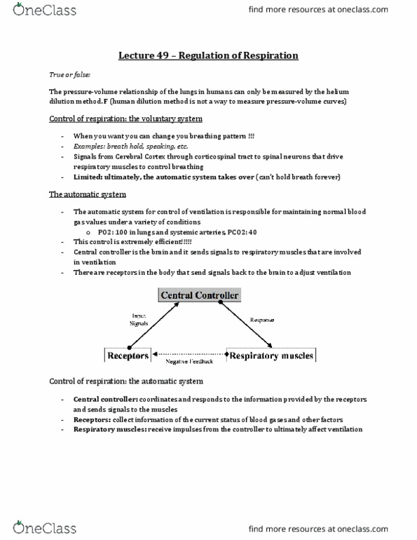
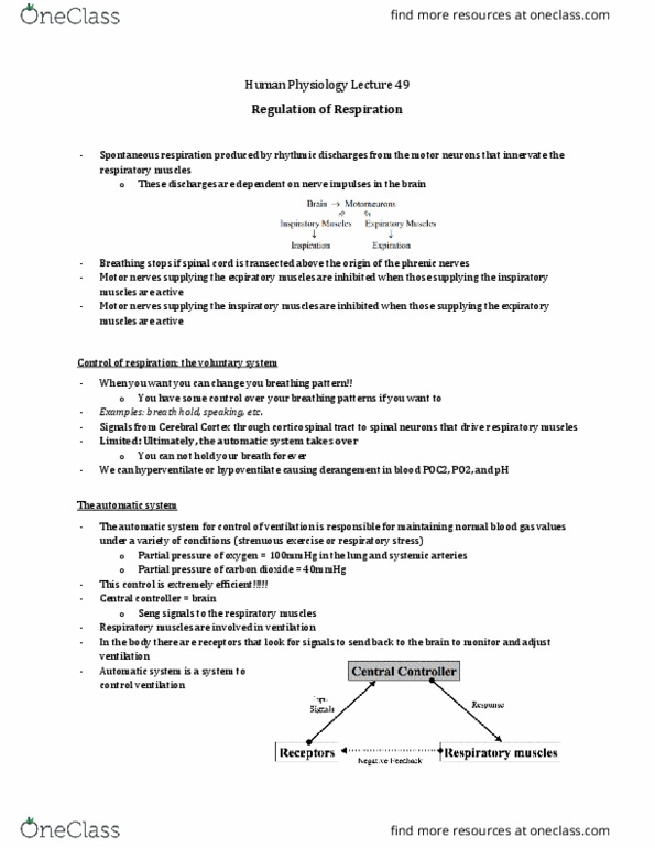
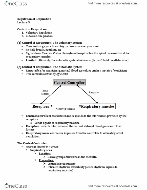
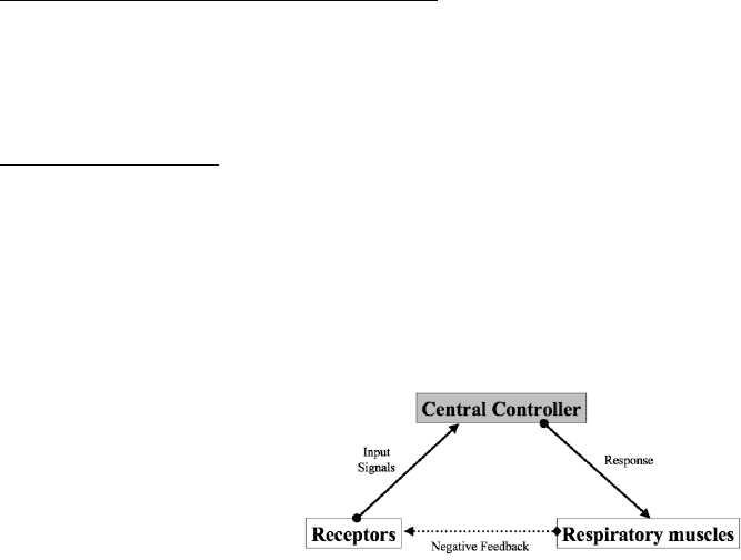
Control of respiration: the automatic system
- Central controller: coordinates and responds to the information provided by the receptors and
sends signals to the muscles
- Receptors: collect information of the current status of blood gases and other factors
- Respiratory muscles: receive impulses from the controller to ultimately affect ventilation
The central controller
- Neurons located in 4 areas
o Inspiratory area
o Pneumotaxic area
o Apneustic center
o Expiratory area
1. Inspiratory area
- Location:
o Dorsal group of neurons in the medulla
- Properties:
o Critical in respiration and sending signals for inspiration
o Most important part for inspiration
o Has rhythmic signals that it sends to the muscles of respiration
o Inherent rhythmic excitability
o Initiates inspiratory drive and ventilation
find more resources at oneclass.com
find more resources at oneclass.com

2. Pneumotaxic area
- Location:
o Group of neurons in the pons
- Properties:
o If there is damage to this area, they take very big breaths
o Fine-tuning of respiration but not essential
o Limits duration of inspiratory drive - switch off
o Control of inspiratory volume and respiratory rate
3. Apneustic center
- Location:
o Group of neurons in the lower pons
- Properties:
o Not sure what it exactly does and if it is really relevant
o opposite of pneumotaxic center in that it can prolong inspiration
o Pneumotaxic center overrides apneustic center
o In healthy situations, this does not contribute to respiration (only if there is damage to
the pneumotaxic center)
4. Expiratory area
- Location:
o Ventral group of neurons in the medulla
- Properties:
o When needed*, these neurons send signals to expiratory muscles resulting in active
expiration
o * (remember expiration is normally a passive process at rest)
Central controller
- Inspiratory area sends signals in regular intervals to muscles to provide regular breathing
- Pneumotaxic area is involved in fine tuning in the system and controls respiratory rate and
volume
- Apneustic center could prolong inspiration if it had a chance but it doesn’t because pneumotaxic
center inhibits it
- Expiratory area allows for active expiration if needed (passive at rest)
Receptors
- Collect information of the current status of blood gases and other factors
- Send signals to controller to change ventilation
- There are 4 types of receptors
o Central chemoreceptors
o Peripheral chemoreceptors
o Lung receptors
o Other receptors
find more resources at oneclass.com
find more resources at oneclass.com
Document Summary
The pressure-volume relationship of the lungs in humans can only be measured by the helium dilution method. F (human dilution method is not a way to measure pressure-volume curves) When you want you can change you breathing pattern !! Signals from cerebral cortex through corticospinal tract to spinal neurons that drive respiratory muscles to control breathing. Limited: ultimately, the automatic system takes over (cid:523)can"t hold breath forever(cid:524) The automatic system for control of ventilation is responsible for maintaining normal blood gas. The automatic system values under a variety of conditions: po2: 100 in lungs and systemic arteries, pco2: 40. Central controller is the brain and it sends signals to respiratory muscles that are involved in ventilation. There are receptors in the body that send signals back to the brain to adjust ventilation. Central controller: coordinates and responds to the information provided by the receptors and sends signals to the muscles.