Physiology 3120 Lecture Notes - Lecture 32: Atrioventricular Node, Pacemaker Potential, Sinoatrial Node

15 May 2018
School
Department
Course
Professor

Lecture 32 - Mechanical performance of the heart
- Changing the force of contraction of the heart is very different than changing the force of
contraction of skeletal muscle (recruit motor units or summate twitch contractions – cannot
be done in the heart)
o There are no motor units in the heart – when one contractile cell has an AP, it
spreads out over all the other contractile cells in the syncytium so it contracts as one
in a wave-like fasion
o Cannot summate the contractile responses of contractile cells because the duration
of the AP is very long (long refractory period)
o = need other ways to change the force of ventricular contraction
Cardiac output
- Cardiac output (CO, Q) is the amount of blood (in liters) pumped by the ventricle in one
minute
- CO is matched to the oxygen demands of the body
o More active = more oxygen cells need
- At rest, oxygen demands are low, and do not need to produce a lot of ATP = CO is low
o CO = 5 L/min (regardless of fitness level)
- Maximal exercise, oxygen demand increases, need lots of ATP, increase in CO
o CO = 20 to 40 l/min (20 – normal, 40 – endurance athlete)
- Stroke volume is the amount of blood ejected from the (left) ventricle when it contracts
(roughly 70 ml at rest)
o EDV - ESV
- CO at rest = 72 bpm x 70 ml = 5 l/min
Cardiac output = heart rate (bpm) x stroke volume
Blood flow distribution under basal (resting) conditions
- Where does the 5L go?
- At rest, a large portion of the blood goes to the:
o Brain: very metabolically active
o Kidneys: filter blood and help maintain pH = receive high amounts of blood
o Liver: largest internal organ in the body = receives high amounts of blood
- Amount of blood muscles receives can change dramatically when the muscles become active
find more resources at oneclass.com
find more resources at oneclass.com

- Cardiac output: depends on heart rate and stroke volume
- Resting CO = 5 l/min → maximum CO = 20 to 40 l/min (depends on level of fitness)
o Normal individual goes from 5 → 20 L/min (mainly by changing HR)
▪ There may be changes in stroke volume for a normal individual but it is not
as dramatic as an endurance athlete)
o Endurance athlete go from 5 → 40 L/min (change HR and SV dramatically)
- Change CO by changing HR and/or SV
Control CO by changes in heart rate
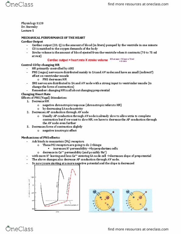
- SA node spontaneously generates APs through changes in principal ions
o Slow depolarization sets up pre-potential to reach threshold and fire AP
- Heart rate is set by the SA node and can be altered by altering the pre-potential:
o 1. Changing the slope of the pre-potential
▪ Increase slope: reach threshold faster, generate APs faster = increase HR
▪ Gradual slope: longer to reach threshold = decrease HR
o 2. Change level of hyperpolarization of membrane potential
▪ Instead of repolarizing to -60 mV, repolarize to -70 mV
▪ = further from threshold = longer to reach threshold
- The factor changing the slope of the pre-potential and the membrane potential is the ANS
- Heart rate is controlled primarily by the autonomic nervous system (ANS)
- SNS (fight or flight) causes increase in heart rate, and therefore, increases cardiac output
- PNS (rest and relax) causes decrease in heart rate, and therefore, decreases cardiac output
Neurotransmitters of the ANS
- ALL preganglionic neurons of both SNS and PNS secrete acetylcholine (Ach – cholinergic
neurons) which binds to nicotinic (N2) receptors in the ganglia or adrenal medulla
- Postganglionic neurons of the PNS also secrete Ach
o Therefore, PNS is totally cholinergic
o Ach binds to muscarinic receptors on target organs (smooth muscle, gland, etc.)
find more resources at oneclass.com
find more resources at oneclass.com

- The receptors are muscarinic and can be blocked by atropine
- Most postganglionic neurons of the SNS secrete norepinephrine (NE) which acts on
adrenergic receptors on target tissues
- Most postganglionic neurons of the SNS are noradrenergic
- The exception to SNS is where:
o Postganglionic neurons of the SNS secrete Ach instead of NE
- These exceptions include:
o 1. Postganglionic sympathetic fibers to sweat glands and piloerector muscles of the
hair secrete Ach
o 2. Postganglionic sympathetic vasodilator nerves innervate blood vessels of skeletal
muscle which secrete Ach (is it in humans?)
- Sympathetic fibers to the adrenal medulla secretes Ach which binds to nicotinic (N2)
receptors to release epinephrine (80%) and norepinephrine (20%)
Controlling CO by changing HR
- Heart rate is primarily controlled by ANS
- PNS (vagus) nerves are distributed mainly to SA and AV nodes and have a small (indirect?)
effect on ventricular muscle cells
o Decrease HR
- SNS nerves are distributed to SA and AV node with a strong input to ventricular muscle
(changing force of contraction)
o Increase HR
- Changing heart = changing slope of pre-potential of SA nodal AP
find more resources at oneclass.com
find more resources at oneclass.com
Document Summary
Lecture 32 - mechanical performance of the heart. Cardiac output (co, q) is the amount of blood (in liters) pumped by the ventricle in one minute. Co is matched to the oxygen demands of the body: more active = more oxygen cells need. At rest, oxygen demands are low, and do not need to produce a lot of atp = co is low. Maximal exercise, oxygen demand increases, need lots of atp, increase in co: co = 5 l/min (regardless of fitness level, co = 20 to 40 l/min (20 normal, 40 endurance athlete) Stroke volume is the amount of blood ejected from the (left) ventricle when it contracts (roughly 70 ml at rest: edv - esv. Co at rest = 72 bpm x 70 ml = 5 l/min. Cardiac output = heart rate (bpm) x stroke volume. Amount of blood muscles receives can change dramatically when the muscles become active. Cardiac output: depends on heart rate and stroke volume.