ANAT-A 215 Lecture Notes - Lecture 6: Pelvis, Renal Corpuscle, Interlobular Arteries

Document Summary
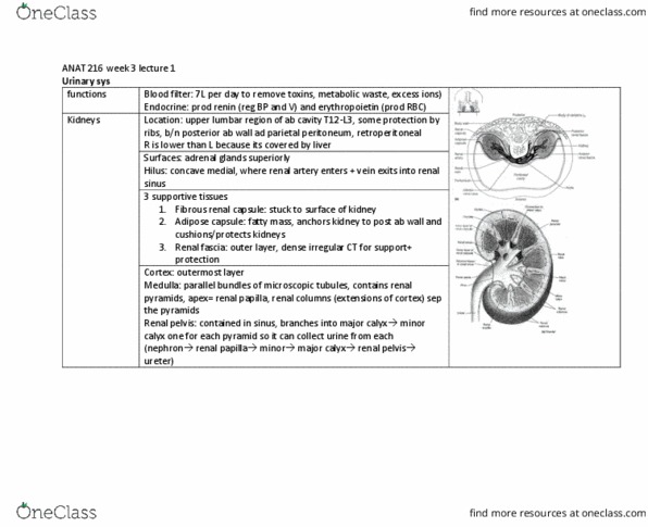
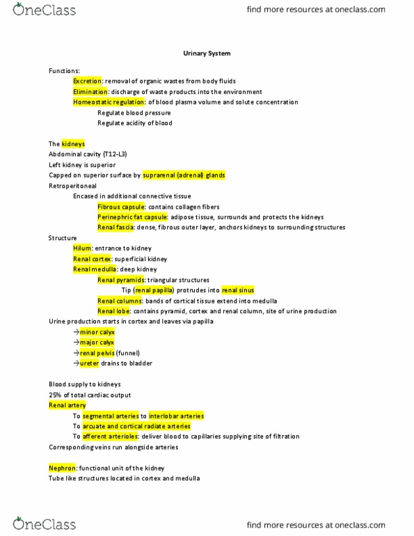
Components (kidneys [2], ureters [2], bladder, urethra: kidneys: gross; external. = innermost perinephric fat (adipose capsule) c. renal fascia, c. t. d. paranephric fat, c. t. = outermost: kidneys: gross; internal, 3 parts, medulla is composed of 8-15 conically-shaped renal pyramids, cortex = vascular; outer, medulla = middle, renal pelvis = inner; tubular area. Cortex contains renal corpuscles (glomerulus [a mass of capillaries] + glomerular [bowman"s] capsule). The filtrate flows into and through the medulla. The pyramids are separated by tissue from the cortex = renal. The apex of the pyramid = renal papillae. The renal papillae are surrounded by funnel-shaped tubes = minor calcyces (sing= calyx) These unite to form the major calyces, which unite to form the renal pelvis (1st part of ureter): renal blood vessels. Some of the plasma (= filtration) leaves the blood stream at this point (fluid = filtrate). Peritubular capillaries primarily in cortex or vasa recta (elongated interlobular v.