Biology 2382B Study Guide - Fluorescence Microscope, Dichroic Filter, Fluorophore

23 Mar 2014
School
Department
Course
Professor
Document Summary
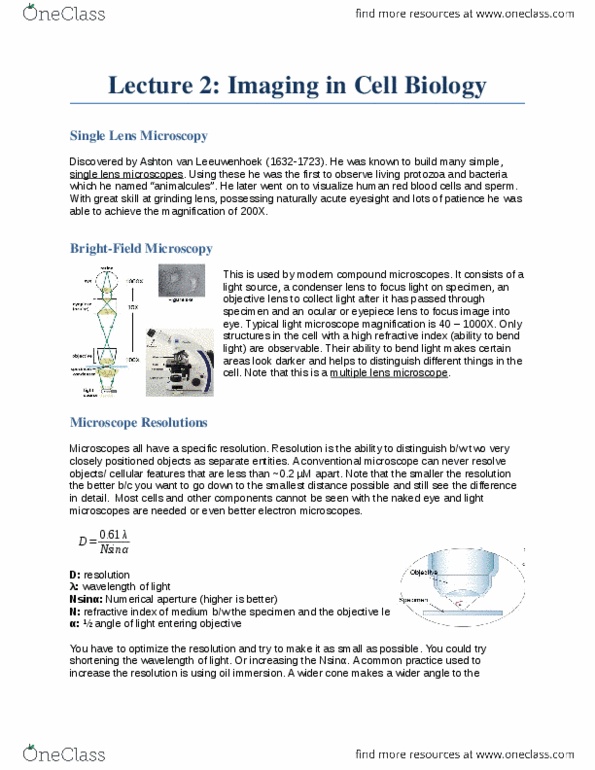
Objective lens to collect light after it has passed through specimen. Ocular or eyepiece lens to focus image onto eye. Typical light microscope magnification is 40 to 1000x. Only structures with a high refractive index (ability to bend light) are observable. Uses visible light to obtain a magnified image. Single cells or thin cell layers but not thick tissues. Location and movement of larger organelles in live cells. Defines outline of large organelles such as nucleus and vacuole and provides better detail of cell edge. Small differences in refractive index and thickness within the cell are further exploited and converted into contrast visible to the eye. Light moves slower in a medium with higher refractive index. Uses a property of certain molecules to fluoresce. Location of fluorescent dyes or fluorescent protein molecules can be imaged. Can visualize more than one protein or cell structure.