BPK 105 Study Guide - Midterm Guide: Intercalated Disc, Skeletal Muscle, Muscle Contraction

27 Apr 2018
School
Department
Course
Professor

Module 3 - Terminology and Objectives - Part 4
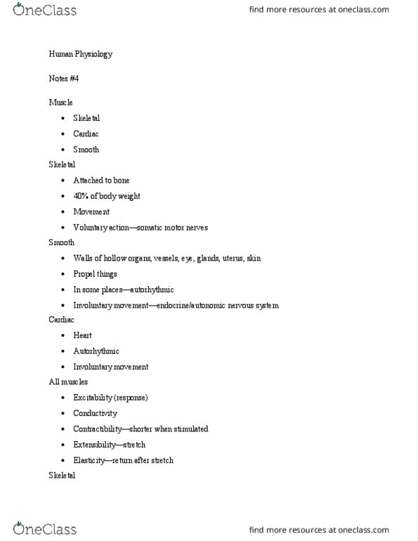
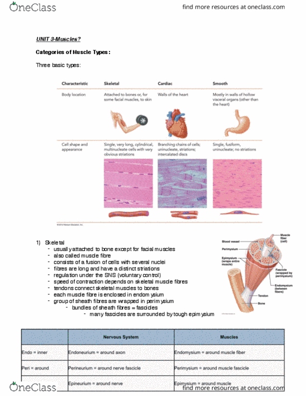
● Differentiate between skeletal, smooth, and cardiac muscles.
SKELETAL
-attached to bone
-long, cylindrical cells
-multiple, peripheral nuclei
-no special features
-has striations (any of the alternating light and dark crossbands that are visible in certain muscle
fibers)
-not autorhythmic
-voluntary control
-moves the whole body
CARDIAC
-heart
-branched cells
-usually single, central nucleus
-special feature: intercalated disks
-has striations
-is autorhythmic
-involuntary control
-contract heart to propel blood through the body
SMOOTH
-walls or hollow organs, blood vessels, and glands
-spindle-shaped cells
-single, central nucleus
-no striations
-is autorhythmic
-involuntary control
-compress organs, ducts, tubes, etc.
Autorhythmic: periodic spontaneous contraction
skeletal- attached to bone, long cylindrical cells, multiple nuclei, striated, voluntary, moves the
body
smooth- located in walls/hollow organs, blood vessels, glands, spindle shaped, single nucleus,
non striated, INvoluntary, compresses organs, ducts, tubes
Document Summary
Module 3 - terminology and objectives - part 4. Differentiate between skeletal, smooth, and cardiac muscles. Has striations (any of the alternating light and dark crossbands that are visible in certain muscle fibers) Contract heart to propel blood through the body. Walls or hollow organs, blood vessels, and glands. Terminology: actin (section 7. 2): a protein that forms (together with myosin) the contractile filaments of muscle cells, and is also involved in motion in other types of cells. 4. myofilaments, extending from z disk to z disk; the structural and functional unit of a muscle. troponin (section 7. 2, figure 7. 9): actin myofilaments, or thin filaments, are made up of three components: actin, troponin, and tropomyosin. The actin strands, which resemble two minute strands of pearls twisted together, have attachment sites for the myosin myofilaments (figure 7. 2 e ). Troponin (tr p -nin) molecules are attached at specific intervals along the actin myofilaments. The end of the axon forms a presynaptic terminal.