ANP 1107 Study Guide - Midterm Guide: Pyelonephritis, Peritoneal Dialysis, Nephron
19 Mar 2019
School
Department
Course
Professor
Document Summary
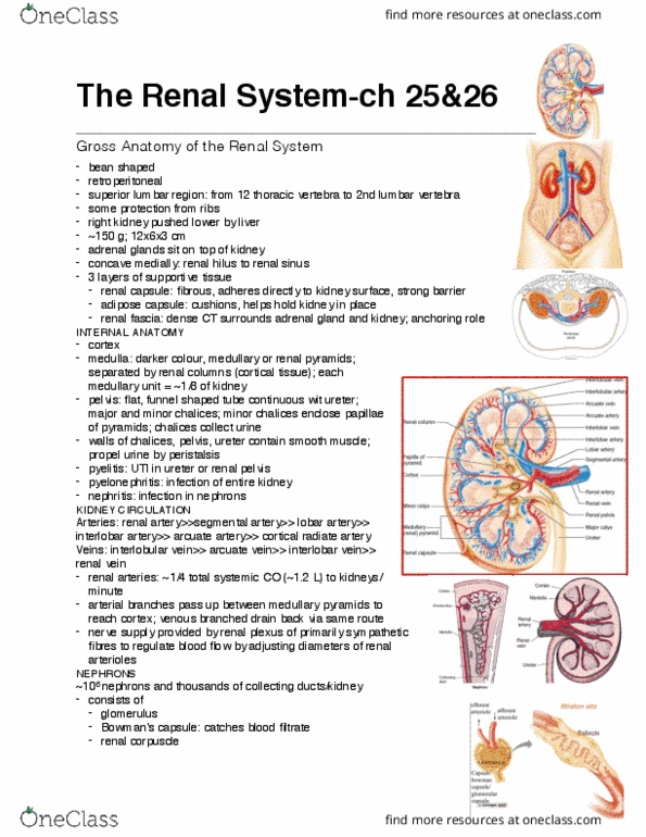
Superior lumbar region, from 12th thoracic vertebrae to. Winter 2019: 3 layers of supportive tissue (from innermost to outermost), renal capsule: fibrous, adheres directly to the kidney surface. Barrier: perirenal fat capsule: acts as a cushion. Helps hold kidney in place: renal fascia: surrounds adrenal gland and kidney; anchors in place. Internal anatomy: cortex: outermost, inner layer after capsule. Blood filtration happens here: medulla: inner area, striped due to urine running. Kidney circulation: renal artery segmental artery interlobar archery arcuate artery cortical radiate artery afferent arteriole afferent arteriole cortical radiate vein arcuate vein interlobar vein renal vein. Nephron: proximal convoluted tubule nephron loop to distal convoluted tubule to collecting duct to papillary duct to minor calyx. collecting duct has: principal cells which lack microvilli and aid in salt and water balance. Intercalated cells have microvilli and regulate acid base balance: 2 types of nephrons, cortical: 85%