Anatomy and Cell Biology 3309 Study Guide - Midterm Guide: Dense Irregular Connective Tissue, Hyaline Cartilage, Loose Connective Tissue

22 May 2018
School
Department
Professor

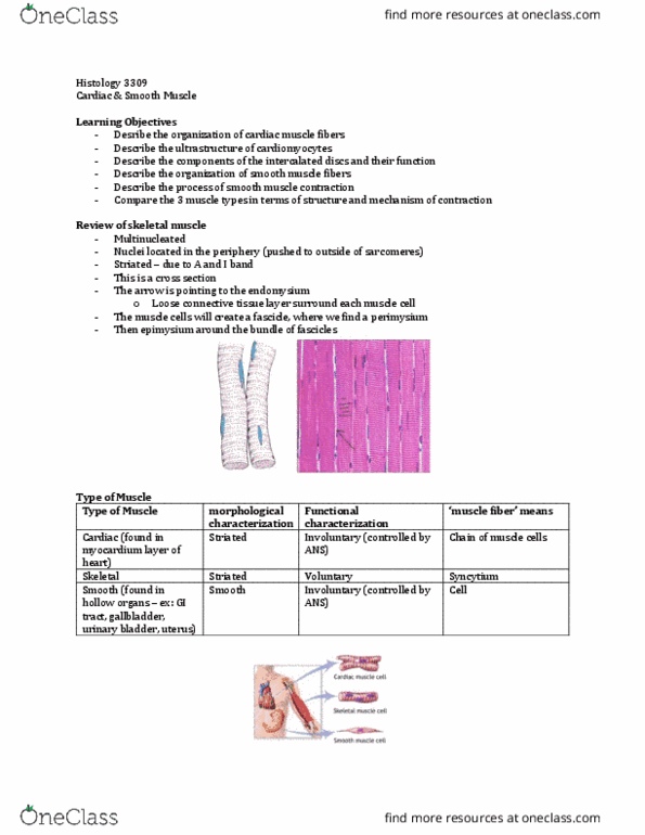
Histology 3309
LAB 5
Classify the tissue
- Dense regular connective tissue
- Only found in tendon and ligament
- Fibers travelling in one direction
- Basophilic cells are fibroblasts
Identify the cell indicated by the yellow arrow
- plasma cells
o Former B lymphocytes that are now activated to create antibodies
o Antibodies bind to the IGe receptors of mast cells
o Has eccentric nucleus
o Has negative golgi (paler staining area in cytoplasm)
o In EM, nucleus would have clock face appearance
Classify the tissue
- loose connective tissue
- more cells than fibers
- the empty space we see here is not empty space its filled with
ground substance
Identify the cell that synthesizes the fibre indicated by the arrow
- yellow arrow is collagen
- the lines are elastin fibers
- the cells are fibroblasts
Cartilage
- 3 types:
o 1) hyaline cartilage
▪ costal cartilage (connects ribs to sternum)
▪ glossy/wet looking
▪ articular cartilage (a type of hyaline cartilage) is found in joints
find more resources at oneclass.com
find more resources at oneclass.com

o 2) elastic cartilage
▪ lots of flexibility and memory
▪ ear
o 3) fibrocartilage
▪ tough
▪ intervertebral disc
• filled with a jelly (rings of fibrocartilage)
▪ pubis symphasis (connection of 2 pubic bones)
General Organization of Cartilage
- perichondrium
o outer most layer
o has 2 layer within it:
▪ fibrous
• dense irregular connective tissue (so collagen fibers are travelling in all
diff directions)
• fibroblasts are in this layer
▪ chondrogenic layer
• thats where you find chondroblasts (build cartilage matrix)
• cells look clumped here
o anytime you see blast it means it produces that cartilage matrix
- chondroblasts undergo appositional growth
o theyre generating so much matrix that they are pushing themselves into the actual
matrix of the cartilage
- once we are in the actual matrix of the cartilage, the cells are called chondrocytes
- chondrocytes undergo interstitial growth
o undergoing mitosis to create daughter cells
- identify by: within cartilage, our cells are arranged in clusters (usually circular) called cell nests
(aka isogenous group)
- usually in hyaline cartilage, we have a ring of deeply staining matrix (called territorial matrix)
around our cell nest
o directly surrounding a cell nest, the GAGs made there are new so they are very
basophilic and they create the territorial matrix
find more resources at oneclass.com
find more resources at oneclass.com
Document Summary
In em, nucleus would have clock face appearance. More cells than fibers the empty space we see here is not empty space its filled with ground substance. Identify the cell that synthesizes the fibre indicated by the arrow yellow arrow is collagen the lines are elastin fibers the cells are fibroblasts. 1: fibrous layer of perichondrium: dense irregular connective tissue. 2: chondrogenic layer of perichondrium: with chondroblasts, more cellular than fibrous layer, cells look plump. 3: new cartilage: when cells are undergoing appositional growth and they first become embedded in cartilage matrix, they don(cid:495)t have that many sulphated gags yet, so its paler at first. Extracellular matrix have hyalonrunoic acid chains branching off them are proteoglycans have core preotein and then branching off of the core protein have sulfated gags very. Reduces friction (mostly function of articular cartilage) Adaptable and pliable tissue: meaning that different daughter cells can adapt to the environment, always undergoing mitosis, forming new cell nests etc.