Anatomy and Cell Biology 3309 Study Guide - Midterm Guide: Haversian Canal, Nuclear Membrane, India Ink

22 May 2018
School
Department
Professor

Lab 7: Central Nervous system
Identify the cell type found within the layer indicated by the box.
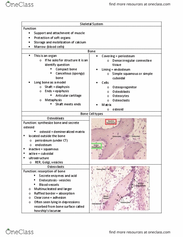
- Osteoblasts (Ob)
o Plump and cuboidal when active
o Undergoing appositional growth to produce lots of matrix (very eosinophilic)
- P = periosteum
o 2 layers: fibrous and osteogenic
Identify the zone indicated by the bracket.
- Zone 2: zone of proliferation
- Above it is zone 1: zone of reserve
o May see isogenous groups
Identify the structure indicated by the arrow.
- Howships lacuna (aka resorption bag)
- Area of bone that has been resorbed (theres no bone left)
- There used to be bone here
- osteoclasts resorbed them
o Osteoclasts are massive and multinucleated
find more resources at oneclass.com
find more resources at oneclass.com

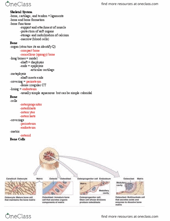
Identify the structure indicated by the arrow.
- This is bone tissue
- The arrow is pointing to lacunae (where an osteocyte would be)
- The dark circle is the haversian canal (where nervous bundle will be)
- This is indian ink (stains spaces not actual cells)
Anatomical Divisions of the Nervous System - CNS
Functional compartments of a neuron
- Owls eye cells
- Soma (cell body) (outlined in red)
- Nucleolus is the dark center and the nucleus is the pale circle around it
- Nucleus is pale bc it is a very active nucleus (tons of euchromatin in there)
- RER and other organelles in the perinuclear space (beside nucleus)
- Nissl bodies
o Darker staining areas in cytoplasm
o Represent the organelles (ex: RER etc) in light microscope
- Axon and dendrites
o Extensions of cell body
o Axon ends in terminal butones
o Nissl bodies extend into the trunk of the dendrites – you wont see them at
the tip of the dendrite extension but you will see darker staining cytoplasmic
components at the base of the dendrites
o Axon hillock – microtubules, neurofilaments but none of the RER →axon
hillock is very pale staining
o Dendrites you will look for nissl bodies extending into the trunk
o And for axon body you will not see anything – very pale staining
find more resources at oneclass.com
find more resources at oneclass.com
Document Summary
Identify the cell type found within the layer indicated by the box. Osteoblasts (ob: plump and cuboidal when active, undergoing appositional growth to produce lots of matrix (very eosinophilic) P = periosteum: 2 layers: fibrous and osteogenic. Above it is zone 1: zone of reserve: may see isogenous groups. Area of bone that has been resorbed (theres no bone left) Osteoclasts resorbed them: osteoclasts are massive and multinucleated. The arrow is pointing to lacunae (where an osteocyte would be) The dark circle is the haversian canal (where nervous bundle will be) This is indian ink (stains spaces not actual cells) Anatomical divisions of the nervous system - cns. Nucleolus is the dark center and the nucleus is the pale circle around it. Nucleus is pale bc it is a very active nucleus (tons of euchromatin in there) Rer and other organelles in the perinuclear space (beside nucleus)