SOSC 1375 Study Guide - Midterm Guide: Palatine Bone, Lambdoid Suture, Articular Processes

7 May 2018
School
Department
Course
Professor

7
Student: _______________________________________________________________________________________
1
.
Which of the followin
g
bones is
p
art of the axial skeleton?
A.
r
ib
B.
r
adius
C. clavicle
D. scapula
E. cox
a
2
.
The a
pp
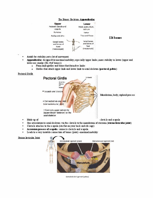
endicular skeleton consists of the
A. skull and appendages
.
B.
r
ib ca
g
e and the
p
elvis
.
C. limbs and their
g
irdles
.
D.
r
ib cage and limb girdles
.
E. vertebral column
.
3
.
Which of the following anatomical features of bones is correctly matched with its function
?
A.
t
ubercle - linin
g
of a
j
oin
t
B.
b
ody - attachment point for a tendon or ligament
C. foramen - a hole for a blood vesse
l
D. sinus - a tunnel in a bon
e
E. foramen - a de
p
ression in a bone
4
.
Which anatomical term is correctl
y
matched with its descri
p
tion
?
A.
c
ondyle - a small, rounded bum
p
B. spine - a low ridge
C.
t
uberosit
y
- a flat, ton
g
ue-sha
p
ed
p
rocess
D.
m
eatus - a tunne
l
E. fossa - adge
Page 1 o
f
49
find more resources at oneclass.com
find more resources at oneclass.com

5
.
Which of the followin
g
bones is
p
art of the neurocranium
?
A.
v
ome
r
B.
n
asal bone
C.
p
alatine bone
D.
o
ccipital bone
E. mandible
6
.
Which of the following bones is paired
?
A.
v
ome
r
B.
t
empora
l
C. s
p
henoi
d
D.
m
andible
E. maxill
a
7
.
Which of the followin
g
bones is a facial bone
?
A.
m
axill
a
B. incus
C.
hy
oi
d
D.
e
thmoi
d
E. s
p
henoi
d
8
.
Which of the following facial bones is correctly matched with its function
?
A.
m
axilla -
p
ossesses sockets for teet
h
B.
v
omer - forms the hard palate
C. inferior nasal conchae - contain nasolacrimal canals
D.
zyg
omatic -
p
rovides attachment
p
oint for tem
p
oralis muscle
E. tem
p
oral - is
p
art of the orbit
.
9
.
A
p
erson who
g
ets hit on the back of the head mi
g
ht suffer in
j
ur
y
to which of these bones
?
A.
t
empora
l
B. occipita
l
C. s
p
henoi
d
D.
z
ygomatic
E. nasa
l
find more resources at oneclass.com
find more resources at oneclass.com

10
.
The sagittal suture is located between the
A.
t
wo
p
arietal bones
.
B. frontal and parietal bones
.
C.
p
arietal and temporal bones
.
D.
p
arietal and occi
p
ital bones
.
E. frontal and temporal bones
.
11
.
The lambdoidal suture is located between the
A.
f
rontal and parietal bones
.
B.
p
arietal and tem
p
oral bones
.
C.
t
em
p
oral and occi
p
ital bones
.
D.
p
arietal and occipital bones
.
E. two parietal bones
.
12
.
The ligamentum nuchae
A.
p
rotects the brain
.
B. is a
p
art of the nose
.
C.
m
oves the eye
.
D. su
pp
orts the
j
aw
.
E. hel
p
s kee
p
the head erect
.
13
.
A person who has cerebrospinal fluid draining from the ear probably has a fracture of the
A.
f
rontal bone
.
B.
t
emporal bone
.
C. z
yg
omatic bone
.
D.
p
arietal bone
.
E. occipital bone
.
14
.
The mastoid
p
rocess
A.
i
s
p
art of the z
yg
omatic arch
.
B. is located anterior to the external auditor
y
meatus
.
C. contains mastoid air cells
.
D.
i
s the point of attachment of the temporalis muscle
.
E. is a sinus
.
15
.
The cheek bone is also known as the
Page 2 o
f
49
find more resources at oneclass.com
find more resources at oneclass.com
Document Summary
What bone does c represent: frontal, maxilla, zygomatic, lacrimal, ethmoid, the figure shows the bones of the right orbit. Page 16 of 49: the figure illustrates the sacrum. What structure does c represent: coccyx, sacral hiatus, superior articular facet, median sacral crest, posterior sacral foramina, the figure illustrates the sacrum. Page 17 of 49: coccyx, sacral hiatus, superior articular facet, median sacral crest, posterior sacral foramina, the figure illustrates the sacrum. What structure does e represent: coccyx, sacral hiatus, superior articular facet, median sacral crest, posterior sacral foramina, the figure illustrates bones of the right upper limb. Page 18 of 49: the figure illustrates bones of the right lower limb. What does b represent: femur, fibula, tarsals, tarsals, tibia, patella, the figure illustrates bones of the right lower limb. What does d represent: femur, fibula, tarsals, tibia, patella, the figure illustrates bones of the right lower limb. What does e represent: femur, fibula, tarsals, tibia, patella.