KAAP220 Chapter Notes - Chapter 9: Myosatellite Cell, Loose Connective Tissue, Muscle Fascicle

28 Nov 2018
School
Department
Course
Professor
Document Summary
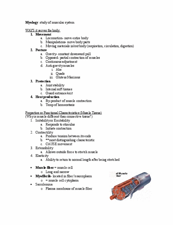
Section 1 - functional anatomy of skeletal muscle tissue. Skeletal muscles: organs composed primarily of skeletal muscle tissue plus connective tissues, nerves, and blood vessels. Epimysium: a dense layer of collagen fibers that surrounds the entire muscle; connected to deep fascia. Muscle fascicle: a bundle of muscle fibers. Perimysium: a fibrous layer that divides the skeletal muscle into a series of compartments. Endomysium: a thin layer of areolar connective tissue that surrounds each muscle fiber. Myosatellite cells: stem cells that function in the repair of damaged muscle tissue. Muscle fibers develop through the fusion of embryonic mesodermal cells called myoblasts. Over time, most of the myoblasts fuse together to form larger multinucleate cells. However, a few myoblasts remain within the tissue as myosatellite cells, even in adults. The multinucleate cells begin differentiating into skeletal muscle fibers as they enlarge and begin producing proteins involved in muscle contraction. Each skeletal muscle fiber has hundreds of nuclei close to plasma membrane.