HTHSCI 1DT3 Lecture Notes - Lecture 17: Foxp3, Epitope, Immunoglobulin A

23 Jun 2018
School
Department
Course
Professor

MHC and TCRs – mark thursz
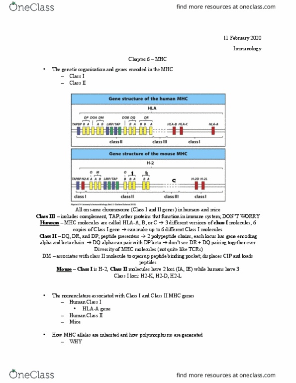
- MHC is on the short arm of chromosome 6
- Some genes are involved in immunology, some aren’t
- TAP and LMP genes are involved in antigen signalling
Class 1 = HLA-A, HLA-B, HLA-C
Class 2 = DP, DM, DQ, DR
Class 3 = TNF-alpha, LTalpha (TNFbeta)
- The allelic diversity is striking; MHC class 1 = 947 alleles (B > A > C)
MHC class 2 = 630 alleles (DRB > DQB > DQA > DP)
- MHC class 1 expressed on all cells except neurones; class 2 only on APCs
- 50, 000 to 100, 000 MHCs are present on a normal cell
- most are occupied by self peptides
- requirements for binding are met by 1/1000 to 1/10 000 random peptides so per cell with all 100000
MHC molecules, the average cell can present around 1000 different peptides
- T cells vary in their threshold requirement for activation, it can be between 2-3 and 500 presenting
MHC molecules
Structure
RESTRICTION is an important concept which describes how T cell responses are restricted for
certain alleles of HLA 1 or HLA 2 AND specific for antigen
HLA structure was shown by mouse MHC type A and targeting it with virus X; the in vitro testing
of CTLs isolated from the mouse’s response then showed they would only clear HLA A cells and
virus X as opposed to other types of virus or HLA molecule
This restriction is because T cells engage variable AAs in the alpha-helix AS WELL AS the correct
specific peptide (antigen) for successful engagement
Both class 1 and 2 MHCs have a transmembrane anchoring region
The MHC CLASS 1 molecule is formed by 2 alpha helices (alpha 2 and alpha 1) which form a cleft,
with a -pleated sheet forming the bottom of the cleft. Underneath these two superior chains
are alpha 3 underneath alpha 2 on the left (an immunoglobulin-like structure which is anchored
to the cell membrane thus stabilises the MHC in the membrane) and 2 macroglobulin
underneath alpha 1 on the right (must be present for MHC molecules to be expressed on cells
hence must be involved in stabilising the structure on the membrane)
The MHC CLASS 2 molecule is formed by 1 alpha helix and one beta-helix which form a cleft,
with a -pleated sheet forming the bottom of the cleft. Underneath these two superior chains
are a2 underneath alpha 1 on the left and 2 underneath 1 on the right
Peptides
Class 1 peptides are 9 aas long; class 2 peptides are less restricted so many 10-11 aas long
Certain aas are commoner depending on the allele due to, these are known as “allele-specific
motifs”
An example of an allele-specific motif is: DRB1*0401 where typically the on of the following aas
will be present in each peptide position:
123456789
W M T L
find more resources at oneclass.com
find more resources at oneclass.com

Y A Q
V S M
L N
Thus in class 1 you can tell from the peptides which MHC side-chain (allelic variant) is present
Antigen processing
CLASS 1: endogenous antigens go to ER (modulated by Transporter Antigen genes (TAP genes)
class 1 molecules loaded with peptide transported to cell surface
CLASS 2: exogenous antigens endocytosed antigens ubiquinated antigens degraded the
proteosome peptide fragments carried to ER via TAP genes meet MHC class 2 molecules in ER
where 2 chains are synthesised: an alpha chain, a beta chain and an invariant chain, the three of
which are complexed together. DM-alpha and DM-beta molecules are chaperone complexes, which
open the closed cleft ends allowing antigen molecules to be loaded into the cleft, depending on
concentration of peptide and affinity of peptide for MHC molecule product transported through
golgi into endosomal compartments, where the invariant chain is degraded by proteases leaving an
alpha chain, beta chain and CLIP protein complex (all that remains of the invariant chain) expressed
on cell surface
N.B. some peptide can enter ER independent of TAP genes
TCRs
Comprised of alpha and beta chains
In gene rearrangement J and V regions are deleted in alpha and beta chain chains, then chains
are translated; the gene rearrangement determines genetic variability which determines which
MHC will be recognised
Pro-thrombocyte gene rearrangement CD4 / CD8 double positive cell selection process in
thymus (positive selection – die by neglect if no affinity for MHC; negative selection – deleted if
affinity for MHC + self)
find more resources at oneclass.com
find more resources at oneclass.com
Document Summary
Mhc is on the short arm of chromosome 6. Some genes are involved in immunology, some aren"t. Tap and lmp genes are involved in antigen signalling. The allelic diversity is striking; mhc class 1 = 947 alleles (b > a > c) Mhc class 2 = 630 alleles (drb > dqb > dqa > dp) Mhc class 1 expressed on all cells except neurones; class 2 only on apcs. 50, 000 to 100, 000 mhcs are present on a normal cell. Requirements for binding are met by 1/1000 to 1/10 000 random peptides so per cell with all 100000. Mhc molecules, the average cell can present around 1000 different peptides. T cells vary in their threshold requirement for activation, it can be between 2-3 and 500 presenting. Restriction is an important concept which describes how t cell responses are restricted for certain alleles of hla 1 or hla 2 and specific for antigen.