HTHSCI 1H06 Lecture Notes - Lecture 2: Tendinitis, Anatomical Terms Of Motion, Aponeurosis

10 Feb 2015
School
Department
Course
Professor


Document Summary
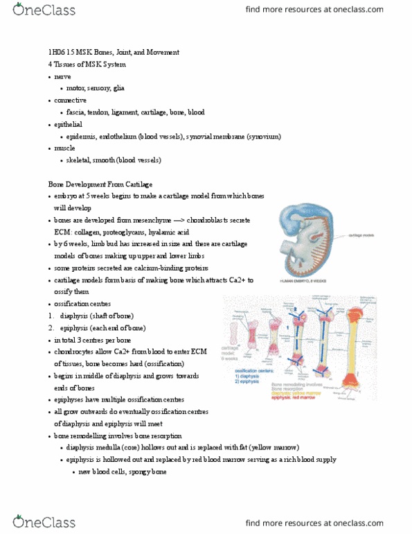
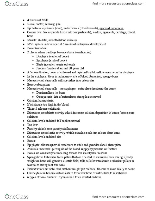
Four tissues of the msk system fascia tendon & ligaments bone & blood: nerve, connective, epithelium epidermis endothelium (blood vessels) synovial membrane, muscle. Bone development from cartilage: starts by condensing one of 3 primary germ tissues (mesenchyme) until it forms cartilage, cartilage cells gather to form models, starts to form connective tissue layer called perichondrium, development of bone within cartilage model forms, develop by calcium carbonate within the bone, cells lay down an ecm made up of minerals. 3 ossification centers: diaphysis: in middle of bone (beginning, epiphysis: at ends of bones (independent) ossification of ecm (cid:224) turns into bone, cartilaginous epiphyseal plate: between the epiphysis and diaphysis, ossification centers don"t fuse until you are 20 inside of bones get hollowed out, in diaphysis, center gets completely hollowed out so we have dense compact bone around outside filled with yellow marrow (mostly fat, in epiphysis, bone hollows itself out in tiny proportions (spongy) filled with red marrow (good blood supply)