BIOM 3200 Lecture 10: 10 Respiratory System

1 May 2018
School
Department
Course
Professor

BIOM3200 – Respiratory System
Pages: 532-574
The Respiratory System
• Divided into respiratory zone (site of gas exchange between air and blood) and a conducting zone
o The exchange of gases between air and blood occurs across the walls of respiratory
alveoli, which permit rapid rates of gas diffusion
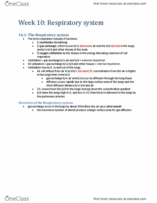
• The term respiration includes three separate (but related) functions:
o Ventilation – breathing
o Gas exchange – between air and blood in lungs; between blood and tissues in body
o Oxygen Utilization – by tissues in energy-liberating reactions of cell respiration
• Ventilation and the exchange of gases between air and blood = external respiration
• Gas exchange between blood and tissues with oxygen utilization = internal respiration
• Ventilation is mechanical process that moves air into and out of the lungs
o Because oxygen concentration of air is higher in lungs than the blood, oxygen diffuses
from the air into blood
o Carbon dioxide moves from the blood to air within lungs by diffusing down its
concentration gradient
o As a result, the inspired air contains more oxygen and less carbon dioxide than expired
air
o Blood leaving the lungs (in pulmonary veins) has a higher oxygen and lower carbon
dioxide concentration than blood delivered to the lungs (in pulmonary arteries)
o The lungs function to bring the blood into gaseous equilibrium with the air
• Gas exchange between the air and blood occurs entirely by diffusion through the lung tissue
o Diffusion occurs very rapidly due to large surface area within the lungs and the small
diffusion distance between blood and air
Structure of the Respiratory System
• Gas exchange in the lungs occurs across ~300 million air sacs = alveoli
o Enormous number provides large surface area (60-80m2) for gas diffusion
o The diffusion rate between the alveolar air and capillary blood depends on distance
separating them
o The thickness of the average alveolar cell and capillary endothelial cells is ~0.15um each,
forming an extremely thin air-blood distance of ~0.3um
• There are two types of alveolar cells:
o Type I alveolar cells
▪ Comprise 95-97% of total surface area of the lungs
▪ Majority of gas exchange with blood occurs here
▪ These cells are very thin (0.15um)
o Type II alveolar cells
▪ Secrete pulmonary surfactant
▪ Reabsorb Na+ and H2O, preventing fluid build up within the alveoli
• In order to maximize the rate of gas diffusion, the air-blood barrier provided by alveoli is
extremely thin with a large surface area
find more resources at oneclass.com
find more resources at oneclass.com

o The alveolar wall is strong enough to withstand high stress during heavy exercise and
high lung inflation
o The great tensile strength of the alveolar wall is provided by the fused basement
membranes (composed of type IV collagen proteins) of the blood capillaries and alveolar
walls
• Alveoli are polyhedral in shape and usually clustered (like units of a honeycomb)
o Air within one member of the cluster can enter the other membranes through tiny pores
o These clusters of alveoli occur of the ends of respiratory bronchioles (very thin air tubes
that end blindly in alveolar sacs)
o Individual alveoli also occur as separate outpouchings along the length of respiratory
bronchioles
o Although the distance between each respiratory bronchiole and terminal alveoli is only
~0.5mm, these units constitute most of the lung mass
• The air passages of the respiratory system are divided into two functional zones:
o Respiratory zone – region where gas exchange occurs (includes respiratory bronchioles
and terminal alveolar sacs)
o Conducting zone – all the of the anatomical structures through which air passes before
reaching respiratory zone (from larynx to terminal bronchioles)
• Air enters the respiratory bronchioles from the terminal bronchioles, which are the narrowest of
the airways that do not have alveoli and do not contribute to gas exchange
o The terminal bronchioles receive air from larger airways formed from successive
branchings of the right and left primary bronchi
o These two large air passages are continuous with the trachea (located in the neck infront
of the esophagus)
o The trachea is a study tube supported by rings of cartilage
• Air enters the trachea from the pharynx (cavity behind the palate that receives the contents of
both the oral and nasal passages)
o In order for air to enter/leave the trachea and lungs, it must pass through valvelike
opening = glottis(between the vocal folds)
o The ventricular and vocal folds are part of the larynx (voice box) which guards the
entrance to the trachea
o The projection at the front of the throat (Adam’s apple) is formed by the largest cartilage
of the larynx
• The conducting zone of the respiratory system consists of the mouth, nose, pharynx, larynx,
trachea, primary bronchi and all successive branchings of the bronchioles up to and including the
terminal bronchioles
o In addition to conducting air into the respiratory zone, these structures serve additional
functions including:
▪ Warming and humidification of inspired air
▪ Filtration
▪ Cleaning
• Regardless of the temperature and humidity of the ambient air, when the inspired air reaches the
respiratory zone, it is at a temperature of 37C (body temp) and is saturated with water vapor as it
flows over the warm, wet mucous membranes that line the respiratory airways
o This ensures that a constant internal body temperature will be maintained while the lung
tissue is protected from desiccation
• Mucous secreted by cells of the conducting zone structures serves to trap small particles in the
inspired air and thereby performs a filtration function
o Mucous is moved along as a rate of 1-2 cm/min by cilia projecting from the tops of
epithelial cells that line the conducting zone
find more resources at oneclass.com
find more resources at oneclass.com

o There are ~300 cilia/cell that beat in coordinated fashion to move mucous towards the
pharynx where it can either be swallowed or expectorated
• As a result of filtration function, particles larger than 6um do not normally enter the respiratory
zone
o Black lung disease in miners who inhaled large amounts of carbon dust from coasl
(causes them to develop pulmonary fibrosis)
o The alveoli themselves are normally kept clean by the action of resident macrophages
o The cleansing action of cilia and macrophages in the lungs is diminished by cigarette
smoke
Thoracic Cavity
• The diaphragm (dome-shaped sheet of striated muscle) divides the anterior body cavity into:
o Abdominopelvic cavity – liver, pancreas, GI tract, spleen, genitourinary tract
o Thoracic cavity – heart, large blood vessels, trachea, esophagus, thymus, lungs
• Structures in the central region (mediastinum) are enveloped by two layers of epithelial
membrane = pleural membranes
o Superficial layer (parietal pleura) lines the inside of the thoracic wall
o Deep layer (visceral pleura) covers surface of the lungs
• The lungs normally fill the thoracic cavity so that the visceral pleura covering each lung is pushed
against the parietal pleura lining the thoracic wall
o Under normal conditions, there is little to no air between viscera and parietal pleura
o There is a potential space (=interpleural space) that can become a real space if the
visceral and parietal pleura separate when lung collapses
Physical Aspects of Ventilation
• The movement of air into and out of the lungs occurs as a result of pressure differences induced
by changes in lung volumes
• Ventilation is influenced by the physical properties of the lungs including their compliance,
elasticity, and surface tension
• Movement of air from higher to lower pressure, between the conducting zone and terminal
bronchioles, occurs as a result of the pressure difference between the two ends of the airway
o Air flow through bronchioles (like blood flow through blood vessels) is directly
proportional to the pressure difference and inversely proportional to the frictional
resistance
o The pressure differences in the pulmonary system are induced by changed in lung
volumes
Intrapulmonary and Intrapleural Pressures
• The visceral and parietal pleurae stick together and the intrapleural space between them contains
a thin layer of fluid (secreted by parietal pleura)
o This fluid is similar to the interstitial fluid is other organs (formed as filtrate from blood
capillaries into the parietal pleura and is drained into lymphatic capillaries)
o The major function of the liquid is to lubricate the lungs so they can slide relative to the
chest during breathing
o Since the lungs are normally stuck to the thoracic wall, they expand and contract with the
wall when breathing
o The intrapleural space is a potential space (becomes real if lungs collapse)
• Air enters the lungs during inspiration because the atmospheric pressure is greater than the
intrapulmonary or intra-alveolar pressure
o A pressure below atmosphere = subatmospheric/negative pressure
find more resources at oneclass.com
find more resources at oneclass.com
Document Summary
Individual alveoli also occur as separate outpouchings along the length of respiratory bronchioles: although the distance between each respiratory bronchiole and terminal alveoli is only. If the baby is premature and lungs have not matured sufficiently to produce surfactant, the effort must be duplicated with every breath. Inspiration and expiration: the diaphragm (innervated by two phrenic nerves composed of axons originating in c3-c5 of the spinal cord) separates the thoracic and abdominal cavities; it is the primary muscle of ventilation. Inspiration is aided by contraction of the parasternal and external intercostals (raise the ribs when they contract and increase thoracic volume laterally: other thoracic muscles become involved in deep inspiration. Pulmonary disorders: asthma, the dyspnea, wheezing and other symptoms of asthma are produced by an onstruction of air flow through the bronchioles that occurs in episodes, this obstruction is caused by inflammation, mucous secretion and bronchoconstriction.