BIOC32H3 Lecture Notes - Lecture 3: Myelin, Axon Hillock, Axon Terminal


LECTURE 3: The Action Potential
Nerve and Muscle Cells are Excitable Cells
● Opening and closing of voltage-gated ion
channels causes changes in membrane
permeability
● Allows ions to move between the IC and
EC compartments
● Leads to changes in excitability of the cell
● In most cells throughout the body,
homeostasis ensures that RMP is
maintained.
○ EXCEPT nerve and muscles cells
which respond to changes in
membrane voltage i.e they are
excitable (ELECTRICAL)
■ muscle contract
■ nerve cells (NEURONS) communicate with each other
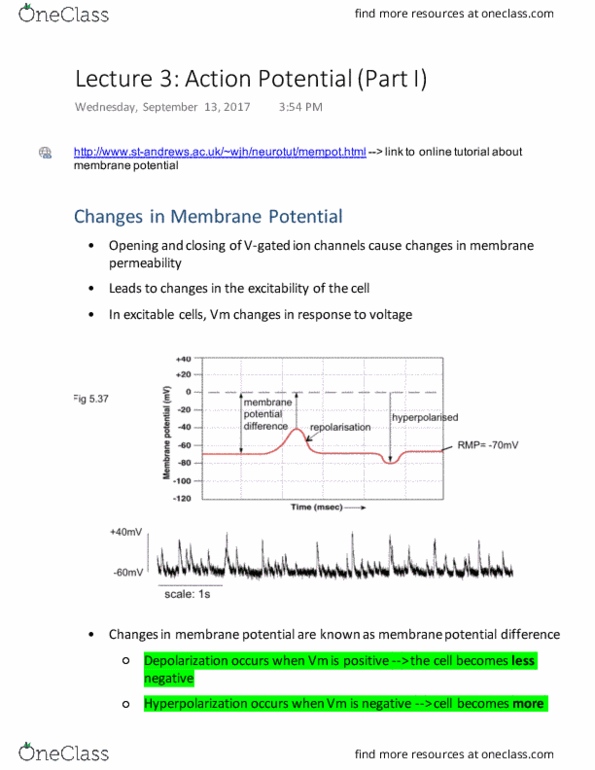
In Excitable Cells Vm changes in Response to Voltage
● Membrane potential difference = Vm in relation to 0
● Towards 0 = depolarisation (less negative)
● More negative than RMP = hyperpolarization
● Towards RMP when depolarised = repolarisation
○ Towards -70 from below
● 2 factors influence a cell’s membrane potential :
○ (1) conc gradients of different ions across the membrane;
○ (2) permeability of the membrane to ions
● If the cell’s permeability to an ion changes, the cell’s membrane potential changes
○ this is an important phenomena when understanding neuronal function
● Here is a trace of an electrical recording. Memb pot begins at rest, i.e -70mV.
○ When the trace moves upwards (ie becomes less negative), the potential difference between inside
and outside the cell is decreased, so the cell is said to be DEPOLARISED or EXCITED.
○ Return to resting Em is known as repolarisation.
○ When the cell moves away form 0mV, ie, becomes more negative, the cell is said to be
HYPERPOLARIZED – essentially more inhibited

Depolarisation is Mainly Due to Opening of Na+ Channels
● Have 2 gates
- only one can be closed at any given time
- open and close rapidly
● During rest, activation gate is closed; sodium channel can become activated (can open and let sodium into
cell)
● Activated when the activation gate is closed
○ when Vm is -55mV or less negative
○ -55 is the threshold
● Whole action potential lasts ~2ms
●Cannot be activated when the inactivation gate is closed
● Na+ ions only flow into the cell when both gates are open
● RMP: activation gate is closed and inactivation gate is open (No Na+ flux)
● Membrane depolarisation, activation gate opens, Na+ ions flow into the cell
● After 0.5milliseconds (ms) inactivation gate closes, preventing more Na+ ion flux
● It takes a further 0.5ms for the gates to reset to default (activation gate is closed and inactivation gate is
open)
● Na and K channels permit movement of ions in both directions
● At rest, activation gate is closed
● Gate opens; NA enters cell , activated
● Structure of sodium channel changes to form a pore where Na can move in
t

There are 2 Types of K+ Channels
1. Leak channels
● Usually open
● Contribute to -70mV resting membrane potential
● Direction of K+ flux depends [K+] in the IC and EC (like diffusion)
2. V-gated K+ channels
● Only one gate to open and close
● Respond to changes in voltage
● Open in response to membrane depolarisation
● Gate is slow to open and close
● Contribute to repolarization and hyperpolarization of the membrane
● Potassium flows slower than sodium
● wBy the time sodium channels close, K is still moving out of the cell → contributes to repolarization
Changes in Membrane Potential Do Not Cause Dramatic Changes in Homeostasis
● Changes in Vm are localised to close to the cell membrane
● Overall no change in charge
● 100mV change in membrane potential: 1 in 100,000 K ions must enter or leave the cell
● The ic and ec composition is relatively stable
Good analogy of this is if you imagine getting sand in your eye and compare it
to all the grains of sand on the beach. You are in a lot of discomfort (big effect
of a few grains), but there is no difference to the beach
The Neuron