PSYB64H3 Lecture Notes - Lecture 2: Ventriculostomy, Ventral Root Of Spinal Nerve, Subarachnoid Space


Chapter 2: Functional Neuroanatomy and the Evolution of the NS
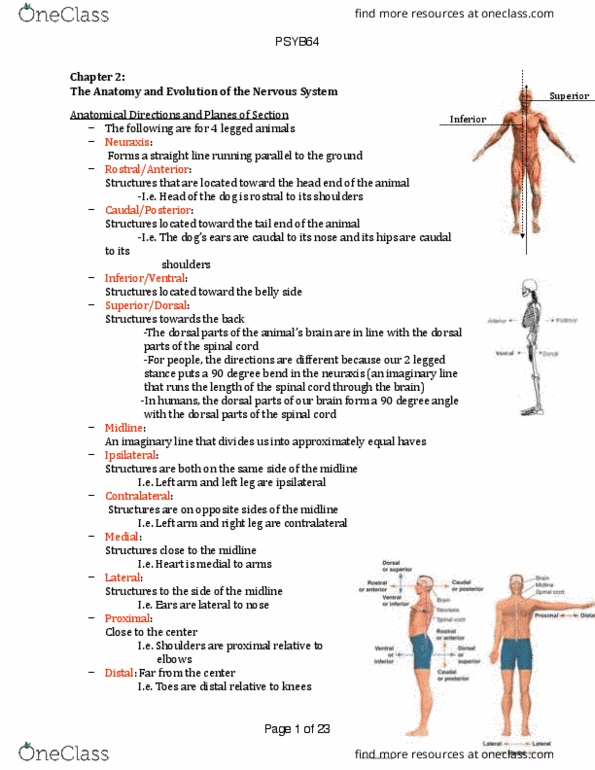
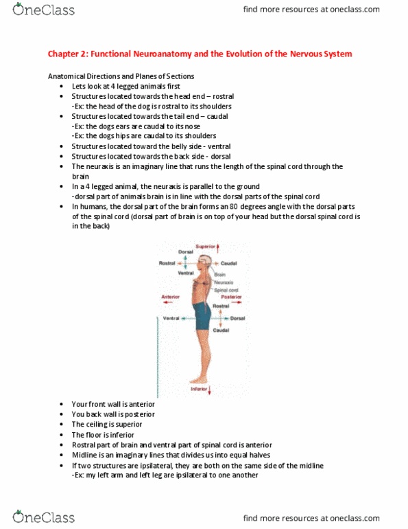
Anatomical Directions and Planes of Section!
1. Rostral: Structures located toward the
head end of the animal !
I) Head of a dog is rostral to its
shoulders !
2. Caudal: Structures located toward the
tail end of the animal !
I) Dogs ears are caudal to its nose &
hips are caudal to its shoulders !
3. Ventral: Structures located toward belly
side !
4. Dorsal: Structure toward the back side!
5. Ceiling= Superior + Floor= Inferior +
Front= Anterior Back= Posterior !
I) Rostral part of the brain and ventral
part of the spinal cord are both
anterior !
6. Midline:!
7. Ipsilateral: (ex. left arm and left leg) vs.
contralateral: (ex. right arm and left
leg) !
8. Medial: Close to midline !
9. Lateral: Structures to the side of the
midline (ex. heart is medial to my arms
whereas my ears are lateral to my
nose) !
10. Proximal: Close to the centre vs. Distal:
Far away from the centre !
I) Refers to limbs (ex. toes are distal relative to my knees and my solders are proximal
relative to my elbows) !
11. 3D as 2D: Sagittal, Midsagittal and horizontal section !

Chapter 2: Functional Neuroanatomy and the Evolution of the NS
!
Protecting and supplying the nervous system !
1. Brain is the most protected organ in the body !
2. Skull is not fully developed in infants—>
Bones overlap each other like tectonic plates !
I) Structure aids in movement of babies
head through birth canal !
II) Soft spot/fontanel: Pulse at the top of the
head between skull bones !
III) Takes 18 months for skull bones to fuse
completely !
!
A. Meninges !
1. Meninges: Layers of membrane that cover
the CNS and the peripheral nerves !
2. Skull to brain layers!
I) Dura mater: Composed of leather like
tissue that follows the outline of the skull
bones !
II) Arachnoid layer: Delicate layer that looks
like a spider web in cross section !
III) Pia mater: Transparent membrane that
sticks closely to the outside of the brain !
3. 3 layers cover the brain and spinal cord but once nerves leave the bony protection of skull
bones and vertebrae of the back= peripheral nerves (protected by dura and pia mater) !
I) Between arachnoid and pia mater is subarachnoid space which contains cerebrospinal
fluid (CSF) !
II) In PNS only the dura mater and pia mater layer cover the nerves (No CSF in PNS)!
III) No arachnoid layer or subarachnoid space surrounding peripheral nerves !
4. Meningitis can result by bacteria and virus attacking meninges layer !
!
B. Cerebrospinal fluid !
1. CSF: Secreted within hollow spaces
in the brain (ventricles) !
I) Within lining of ventricle, choroid
plexus converts material in blood
supply—> CSF !
II) CSF composition: Similar to
composition of clear plasma in
blood!
III) Floats the brain within the skull !
2. Advantages: !
I) Acts as a cushion!
II) Bump head—> neurone respond
to appropriate input not to
pressure the brain !
3. Pressure can cause neurone to fire
(ex. tumor causes seizures by
pressing down a part of the brain!

Chapter 2: Functional Neuroanatomy and the Evolution of the NS
4. Circulates through the central canal of the spinal cord and 4 ventricles in the brain !
I) Two lateral ventricles (one in each hemisphere) !
II) 3rd and 4th ventricle in the brainstem !
III) 4th ventricle is continuous with central canal of the spinal cord which runs the length of
the cord at its midline !
5. Below the 4th ventricle there is a small opening to allow the CSF to flow into the
subarachnoid space that surrounds the brain and spinal cord. !
6. Entire supply of CSF is turned over 3x per day!
I) Old supply is reabsorbed into blood supply at the top of the head !
7. Production & Exit !
I) Produced by the choroid plexus (lines the wall of ventricle) !
II) 2 Lateral ventricle—> 3rd and 4th ventricle—> central canal (spinal cord)!
III) Exit: Base of cerebellum into the subarachnoid space—> Reabsorbed by veins near top
of the head!
8. Blockages= Hydrocephalus (water on the head)!
I) Occurs in birth !
II) Diagnosing: Neurological exam, prenatal
exam, and brain imaging !
III) Left untreated—> intellectual disability (large
quantity of CSF prevents normal growth of
brain) !
IV) Treatment: Insulation of shunt in one ventricle
of the brain to drain off excess fluid to
abdomen or heart or ventriculostomy which
involves making a hole in the third ventricle or
between 2 ventricle to improve circulation of
CSF !
V) In adults: Tumors or scar tissue can cause
blockage of CSF and can be treated w/ shunts
and/or surgery !
9. Spinal tap: Fluid is withdrawn from subarachnoid
space with a needle to diagnose diseases !
I) CSF moves through a separate circulation
system than blood !
!
C. The brain blood supply !
1. 15-20% of blood is pumped to the brain !
2. Arteries!
I) Carotid arteries: 1/2 major blood vessels
that travel up the sides of the neck to supply
the brain !
II) Vertebral arteries: 1/2 major blood vessel
that enters the brain from the back of the
skull!
3. Once in the skull the major arteries form the
anterior, middle, and posterior cerebral arteries
which serve most of the brain !
4. Interruption of blood supply—> Significant
damage of the brain in less than 3 minutes !
Document Summary
Front= anterior back= posterior : rostral part of the brain and ventral part of the spinal cord are both anterior , midline:! Far away from the centre : refers to limbs (ex. toes are distal relative to my knees and my solders are proximal relative to my elbows) , 3d as 2d: sagittal, midsagittal and horizontal section ! Protecting and supplying the nervous system : brain is the most protected organ in the body , skull is not fully developed in infants > Bones overlap each other like tectonic plates : structure aids in movement of babies head through birth canal ! Ii) soft spot/fontanel: pulse at the top of the head between skull bones ! Ii) arachnoid layer: delicate layer that looks like a spider web in cross section ! In pns only the dura mater and pia mater layer cover the nerves (no csf in pns)! Ii) csf composition: similar to composition of clear plasma in blood!