Anatomy and Cell Biology 3309 Lecture Notes - Lecture 11: Lymph Node, Mantle Zone, B Cell

22 May 2018
School
Department
Professor

Histology 3309
Lymph Nodes
Question
Which of the following is a way in which lymphatic vessels are DIFFERENT from veins?
a) Only lymphatic vessesl carry lymphocytes
b) Lymphatic vessels have anchoring filaments
c) Lymphatic vessles have valves
d) Lymphatic vessels only move fluid in one direction
A: B
Rupert Mclean Case Study
- They were really concerned with metastasis
- If the cancer is localized, some cancers are very treatable (this is the case with melanoma)
- But bc Rupert had this paint splatter pattern it wasnt just one mole they were really concered
with metastasis
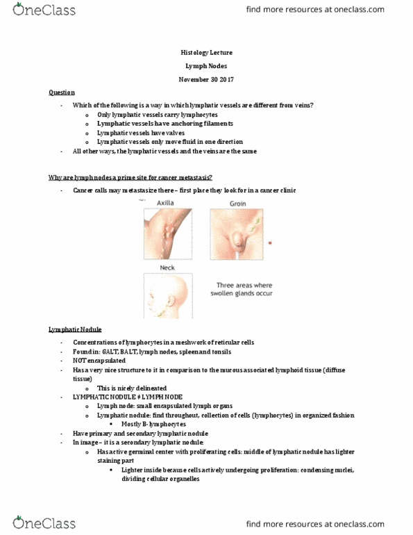
Why are lymph nodes a prime site for cancer metastasis?
- They are concentrated in diff areas: axilla, groin and neck
Lymphatic Nodule
- Concentrations of lymphocytes in a meshwork of reticular cells
o Primarily B lymphocytes that make up lymphatic nodule
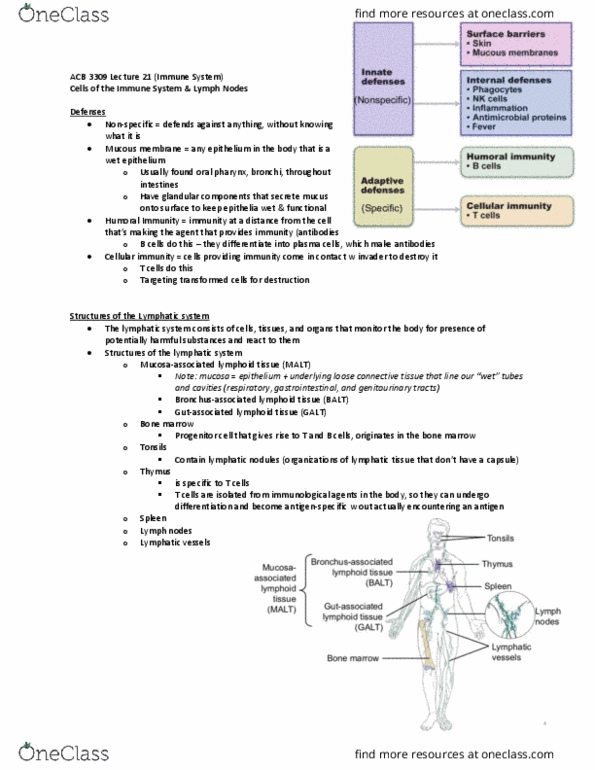
- Found in:
o MALT: GALT & BALT
o Lymph nodes
o Spleen
o Tonsils (this is a collection of lymphatic nodules)
- NOT encapsulated
- The entire blue is a lymphatic nodule (LN)
- Very nice overall structure to it in comparison to the other mucosa associated lymphatic tissue
(MALT, BALT or GALT) which is kinda diffuse
- Difference bw lymphatic nodule and lymph node:
o Lymphatic nodule is a collection of cells in an organized fashion you can find in your
body
o Lymph nodes are small encapsulated, bean shaped lymph organs in our body
- This image is a secondary lymphatic nodule
- secondary lymphatic nodules have active germinal center with proliferating cells
o in the middle there is a lighter staining part where cells are actively undergoing
proliferation
o they are condensing their nuclei, dividing their cellular organelles and proliferating
find more resources at oneclass.com
find more resources at oneclass.com

o whereas in the outside (called the mantle zone), the cells are mostly B lymphocytes that
are not actively proliferating (so very basophilic staining)
- when lymphocytes are in circulation, they are basically just nuclei (their nucleus takes up most
of the cell)
- when they are proliferating, they are going to be dividing their contents so we dont have that
super dark concentration of staining of nuclei from the lymphocytes
- this image is in the intestinal gland
Primary VS Secondary Lymphatic Nodules
Primary
Secondary
No germinal centre only mantle zone
Germinal AND mantle zones
Lymphocytes not yet activated
Lymphocytes activated (proliferation) so these
have encountered their antigen
Few (not going to see a lot of these when looking
in microscope)
Numerous (will see a lot of these)
find more resources at oneclass.com
find more resources at oneclass.com

Objectives
- Describe the structure of the lymph node
- Explain the path of lymph flow to and from the lymph node
- Evaluate the role of the supporting elements of the lymph node to the overall structure of the
lymph node
- Compare and contrast the structure, appearance, function and cell composition of the cortex and
medulla
- Describe the route of lymph flow through the sinuses of the lymph node
Functions of the Lymph Node
- Involved in filtration
o We have lots of lymphocytes in the lymph node with all these immune cells contained
within filtering that lymph – picking up anything unwanted
- Involved in interaction
o Activation
- Differentiation of cells
o B cells can differentiate into either memory cell or plasma cell
o Helper T cells are important for that
o T cells can also differentiate
- Lymph nodes are working all the time (get swollen all the time – even when you are not sick)
Remember: Primary VS Secondary Lymphoid Organs
- When we are talking about lymph nodes, we are talking about them as secondary lymphoid
organs
- This is the battle ground – where cells launch an immune response
- Cells get activated, we have effector and memory cells that become able to go out and fight the
infection
- Recall: vaccination – we are priming the cells, the first time around, B cells are activated and
they make memory cells so the next time we encounter the pathogen, they are primed and ready
to attack it
find more resources at oneclass.com
find more resources at oneclass.com
Document Summary
Which of the following is a way in which lymphatic vessels are different from veins: only lymphatic vessesl carry lymphocytes, lymphatic vessels have anchoring filaments, lymphatic vessles have valves, lymphatic vessels only move fluid in one direction. If the cancer is localized, some cancers are very treatable (this is the case with melanoma) But bc rupert had this paint splatter pattern (cid:523)it wasn(cid:495)t just one mole(cid:524) they were really concered with metastasis. They are concentrated in diff areas: axilla, groin and neck. Concentrations of lymphocytes in a meshwork of reticular cells: primarily b lymphocytes that make up lymphatic nodule. Found in: malt: galt & balt, lymph nodes, spleen, tonsils (this is a collection of lymphatic nodules) The entire blue is a lymphatic nodule (ln) Very nice overall structure to it in comparison to the other mucosa associated lymphatic tissue (malt, balt or galt) which is kinda diffuse.