Anatomy and Cell Biology 3309 Lecture Notes - Lecture 1: Corneal Epithelium, Stroma Of Cornea, Corneal Endothelium

29 Apr 2021
School
Department
Professor
Document Summary
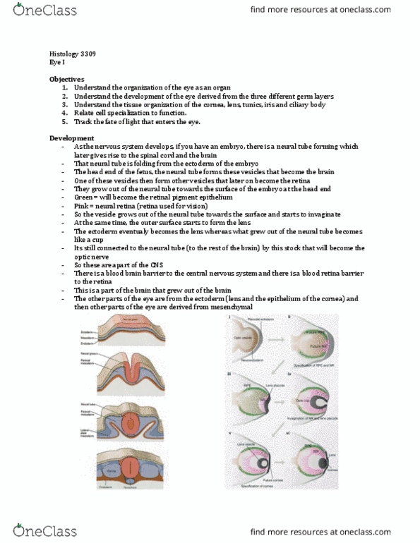
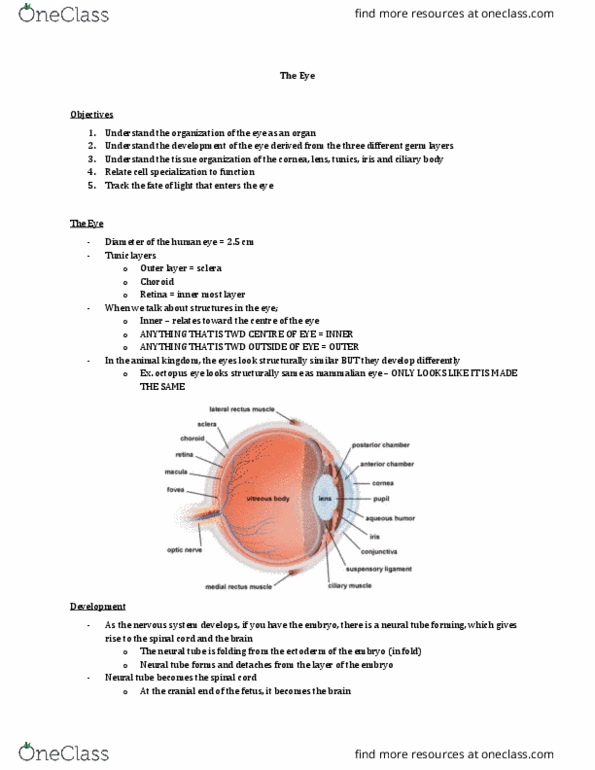
Derived from neuroectoderm, surface ectoderm and mesoderm. As neural tube forms, optical vesicles are formed at cranial end. Connection to forebrain constricts optic stalk. Contact to surface ectoderm induces lens placode. Invagination of optic vesicle & lens placode forms optic cup with retina and retinal pigment epithelium (rpe) plus lens vesicle. Neuroectoderm: retina, rpe, iris, ciliary body epithelium. Mesoderm: corneal endothelium & stroma, sclera, choroid, blood vessels, etc. Sclera, (hard), the white of the eye, opaque, often blue in kids and yellow in elderly. Dark brown due to melanocytes and blood vessels. Ciliary body: smooth muscle (radial and concentric) lens accomodation. Iris: smooth muscles (radial and concentric) aperture: pupil (black) light adaption. Thin layer of two components: neural retina and rpe. Anterior and posterior contain aqueous humor, vitreous chamber contains vitreous humor. Light passes through cornea, anterior chamber, pupil, posterior chamber, lens, vitreous chamber to the retina. The main refraction is provided by the cornea.