Biology 2382B Lecture Notes - Lecture 3: Hoechst Stain, Differential Centrifugation, Centrifugal Force

16 Aug 2017
School
Department
Course
Professor
Document Summary
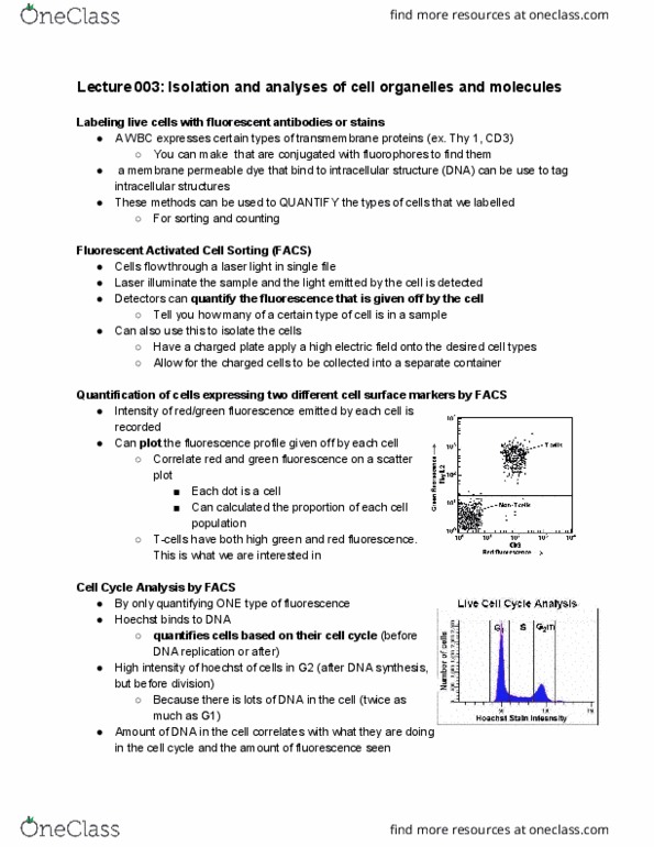
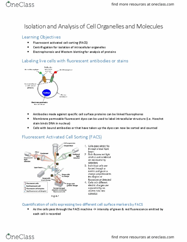
Isolation and analysis of cell organelles and molecules. Electrophoresis and western blotting for analysis of proteins. Labelling live cells with fluorescent antibodies or stains. Antibodies made against specific cell surface proteins can be linked to fluorophores: wbc express cd3 and thy1. 2 cell surface proteins, note: fluorophore-linked secondary antibodies could also be used in combination with unlabeled primary antibodies. Membrane permeable fluorescent dyes can be used to label intracellular structures (i. e. hoechst stain binds dna in nucleus: make sure that these are nucleated cells. Cells with bound antibodies or that have taken up the dyes can now be sorted and counted. Fluorescent activated cell sorting (facs: cells pass single file through a laser light beam, both fluorescent light emitted and scattered are measured by detectors. Quantification of the amount of fluorescence: all cells passing through should be blue because they are nucleated. Quantification of cells expressing two different cell surface markers by facs.