Biology 2382B Lecture Notes - Lecture 7: P53, Fas Ligand, Cytokine

23 Aug 2016
School
Department
Course
Professor
Document Summary
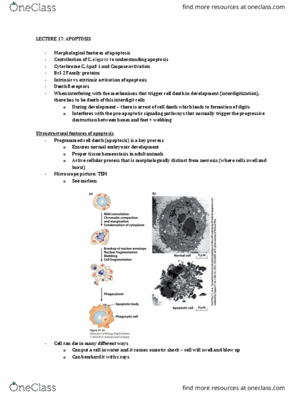
Ultrastructural features of apoptosis programmed cell death (apoptosis) is a key process: ensures normal embryonic development, proper tissue homeostasis in adult animals, active cellular process that is morphologically distinct from necrosis. 3 researchers won nobel prize in physiology (2002) for their discoveries concerning genetic regulation of organ development and programmed cell death: sydney brenner, robert horvitz, john sulstan. Advantages of studying c. elegans: small (1mm long, transparent, every single cell has been mapped out. 959 cells in adult hermaphrodites, 1031 in adult males: genome fully sequenced, many genetic mutants. Mutations in ced-3 gene block apoptosis in c. elegans out of 1090 newborn cells, 131 die during development, resulting in a nematode with 959 cells dead cells are highly refractile and can be detected by dic microscopy. Caspase cysteine-dependent aspartate-directed proteases two types: initiator caspases: cleave inactive pro-forms of effector caspases activates them, effector (executioner) caspases: cleave other protein substrates within the cell triggers apoptotic process.