Physiology 3140A Lecture Notes - Lecture 22: Transducin, Cell Membrane, Protein Kinase

28 Dec 2017
School
Department
Course
Professor
Document Summary
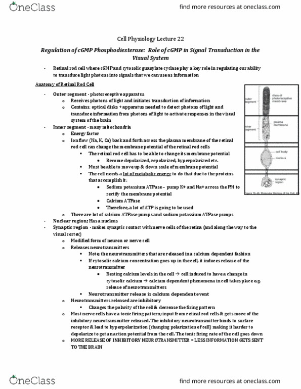
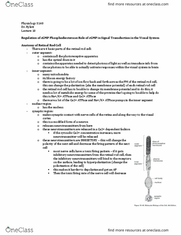
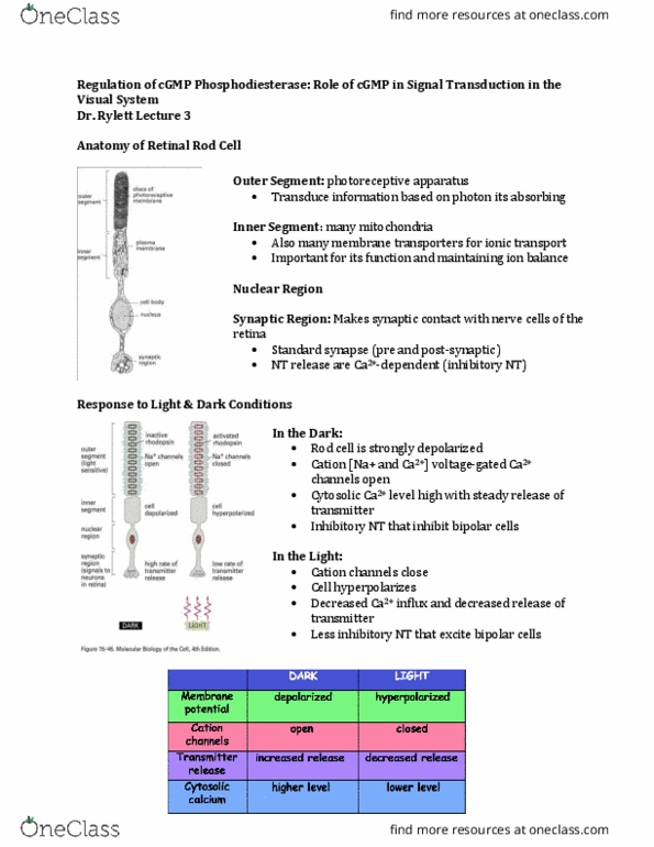
Role of cgmp in signal transduction in visual system. Inner segment: many mitochondria: energy factory, many ions flow back and forth across pm of retinal rod cell (sodium, calcium, potassium) Ion flow can change the polarization/membrane potential or retinal rod cells: rod cell must be able to change its membrane potential. If it becomes depolarized/hyperpolarized, it must repolarize: needs a lot of metabolic energy because the proteins that accomplish the repolarization are, sodium/potassium atpase, calcium-atpase pumps, there are many sodium/potassium atpases and calcium-atpases located here. Synaptic region: makes synaptic contact with nerve cells of the retina to the visual cortex, modified nerve cell releases inhibitory neurotransmitters at the synaptic region, neurotransmitter is released in a calcium-dependent fashion. Most nerve cells have a tonic firing pattern. It is the next cell in line after retinal rod cells: retinal rod cell releases more inhibitory neurotransmitter on the nerve cell.